

Proteomic analysis of human periodontal ligament cells under hypoxia
- PMID: 31496921
- PMCID: PMC6717648
- DOI: 10.1186/s12953-019-0151-2
Proteomic analysis of human periodontal ligament cells under hypoxia
Abstract
Background: The periodontal ligament is essential for homeostasis of periodontal tissue. A hypoxic milieu of the periodontal tissue is generated under periodontitis or during orthodontic treatment, which affects the periodontal and bone remodelling process. Here, we provide a comprehensive proteomic characterization of periodontal ligament cells under hypoxic conditions, aiming to reveal previously unappreciated biological changes and to help advance hypoxia-based therapeutic strategies for periodontal diseases.
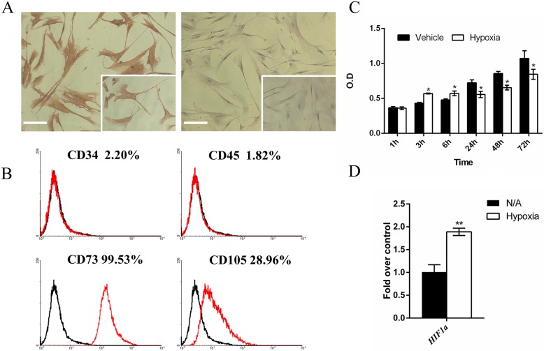
Methods: Human periodontal ligament cells (hPDLCs) were characterized using immunohistochemistry (IHC) and flow cytometry (FACS). Successful hypoxia treatment of hPDLCs with 1% O2 was confirmed by qRT-PCR. Proliferation was evaluated using an MTT assay. The proteomic expression profile under hypoxia was studied with the isobaric tags for relative and absolute quantification (iTRAQ) approach followed by protein identification and bioinformatic analysis, and western blot verification was performed.
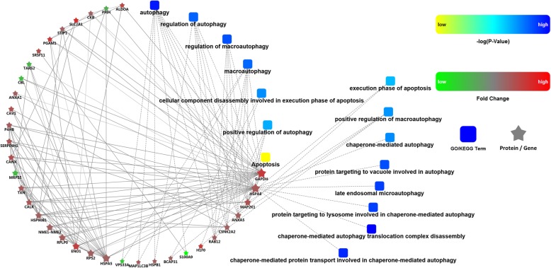
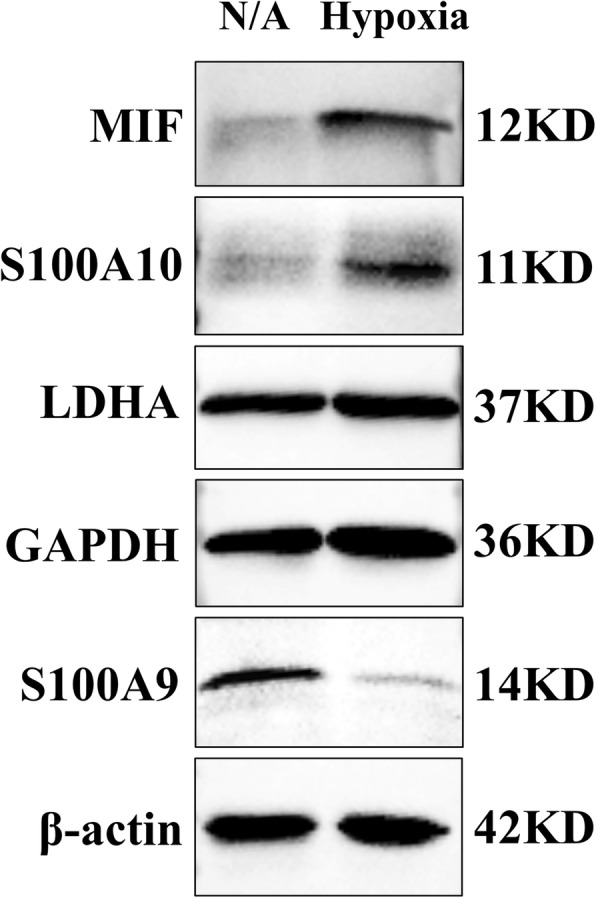
Results: The hPDLCs were positive for vimentin, CD73 and CD105 and negative for keratin, CD34 and CD45. After hypoxia treatment, the mRNA expression of hypoxia-inducible factor 1a (HIF1a) was upregulated. The proliferation rate was elevated during the first 6 h but decreased from 6 h to 72 h. A total of 220 differentially expressed proteins were quantified in hPDLCs under hypoxia (1% O2, 24 h), including 153 upregulated and 67 downregulated proteins, five of which were verified by western blot analysis. The Gene Ontology enriched terms included the energy metabolic process, membrane-bound organelle and vesicle, and protein binding terms. Kyoto Encyclopedia of Genes and Genomes (KEGG) analysis indicated several involved pathways, including glycolysis/gluconeogenesis, biosynthesis of amino acids, the HIF-1 signalling pathway, and focal adhesion. A protein-protein interaction (PPI) network demonstrated the dominant role of autophagy over apoptosis under hypoxia.
Conclusion: The proteomic profile of hPDLCs under hypoxia was mainly related to energy metabolism, autophagy, and responses to stimuli such as adhesion and inflammation. Previously unrecognized proteins including solute carrier family proteins, heat shock proteins, ubiquitination-related enzymes, collagen and S100 family proteins are involved in adaptive response to hypoxia in hPDLCs and are thus of great research interest in future work.
Keywords: Fibroblasts; Hypoxia; Pathogenesis of periodontal disease(s); Periodontal ligament; Proteome.
Conflict of interest statement
Competing interestsThe authors declare that they no competing interests.
Figures


Similar articles
-
Hypoxia-regulated human periodontal ligament cells via Wnt/β-catenin signaling pathway.Medicine (Baltimore). 2017 Apr;96(16):e6562. doi: 10.1097/MD.0000000000006562. Medicine (Baltimore). 2017. PMID: 28422843 Free PMC article.
-
Leptin attenuates hypoxia-induced apoptosis in human periodontal ligament cells via the reactive oxygen species-hypoxia-inducible factor-1α pathway.Exp Physiol. 2021 Aug;106(8):1752-1761. doi: 10.1113/EP089324. Epub 2021 Jul 6. Exp Physiol. 2021. PMID: 34143536
-
Comparative proteomic profiling of human dental pulp stem cells and periodontal ligament stem cells under in vitro osteogenic induction.Arch Oral Biol. 2018 May;89:9-19. doi: 10.1016/j.archoralbio.2018.01.015. Epub 2018 Feb 3. Arch Oral Biol. 2018. PMID: 29407636
-
Evaluation of Hypoxia on the Expression of miR-646/IGF-1 Signaling in Human Periodontal Ligament Cells (hPDLCs).Med Sci Monit. 2018 Jul 30;24:5282-5291. doi: 10.12659/MSM.910163. Med Sci Monit. 2018. PMID: 30058629 Free PMC article.
-
Enamel matrix proteins regulate hypoxia-induced cellular biobehavior and osteogenic differentiation in human periodontal ligament cells.Biotech Histochem. 2017;92(8):606-618. doi: 10.1080/10520295.2017.1370131. Epub 2017 Dec 5. Biotech Histochem. 2017. PMID: 29205072
Cited by
-
The effect of hypoxia on the proteomic signature of pig adipose-derived stromal/stem cells (pASCs).Sci Rep. 2020 Nov 18;10(1):20035. doi: 10.1038/s41598-020-76796-7. Sci Rep. 2020. PMID: 33208768 Free PMC article.
-
Research progress of hydrogel therapy to improve hypoxic environment of periodontal tissues and promote periodontal regeneration.J Oral Biol Craniofac Res. 2025 Jul-Aug;15(4):869-879. doi: 10.1016/j.jobcr.2025.06.005. Epub 2025 Jun 13. J Oral Biol Craniofac Res. 2025. PMID: 40586108 Free PMC article. Review.
-
Long non-coding RNA 01126 promotes periodontitis pathogenesis of human periodontal ligament cells via miR-518a-5p/HIF-1α/MAPK pathway.Cell Prolif. 2021 Jan;54(1):e12957. doi: 10.1111/cpr.12957. Epub 2020 Nov 24. Cell Prolif. 2021. PMID: 33231338 Free PMC article.
-
Mediating and moderating effects of plasma proteomic biomarkers on the association between poor oral health problems and incident dementia: The UK Biobank study.Geroscience. 2024 Oct;46(5):5343-5363. doi: 10.1007/s11357-024-01202-3. Epub 2024 May 29. Geroscience. 2024. PMID: 38809392 Free PMC article.
-
Targeted proteomics addresses selectivity and complexity of protein degradation by autophagy.Autophagy. 2025 Feb;21(2):460-475. doi: 10.1080/15548627.2024.2396792. Epub 2024 Sep 20. Autophagy. 2025. PMID: 39245437 Free PMC article.
References
LinkOut - more resources
Full Text Sources
Research Materials
Miscellaneous
