. 2019 Aug 21:10:1100.
doi: 10.3389/fphys.2019.01100. eCollection 2019.

Nociceptive Pathway in the Cockroach Periplaneta americana

Affiliations

Nociceptive Pathway in the Cockroach Periplaneta americana

Stav Emanuel et al. Front Physiol. .

Abstract

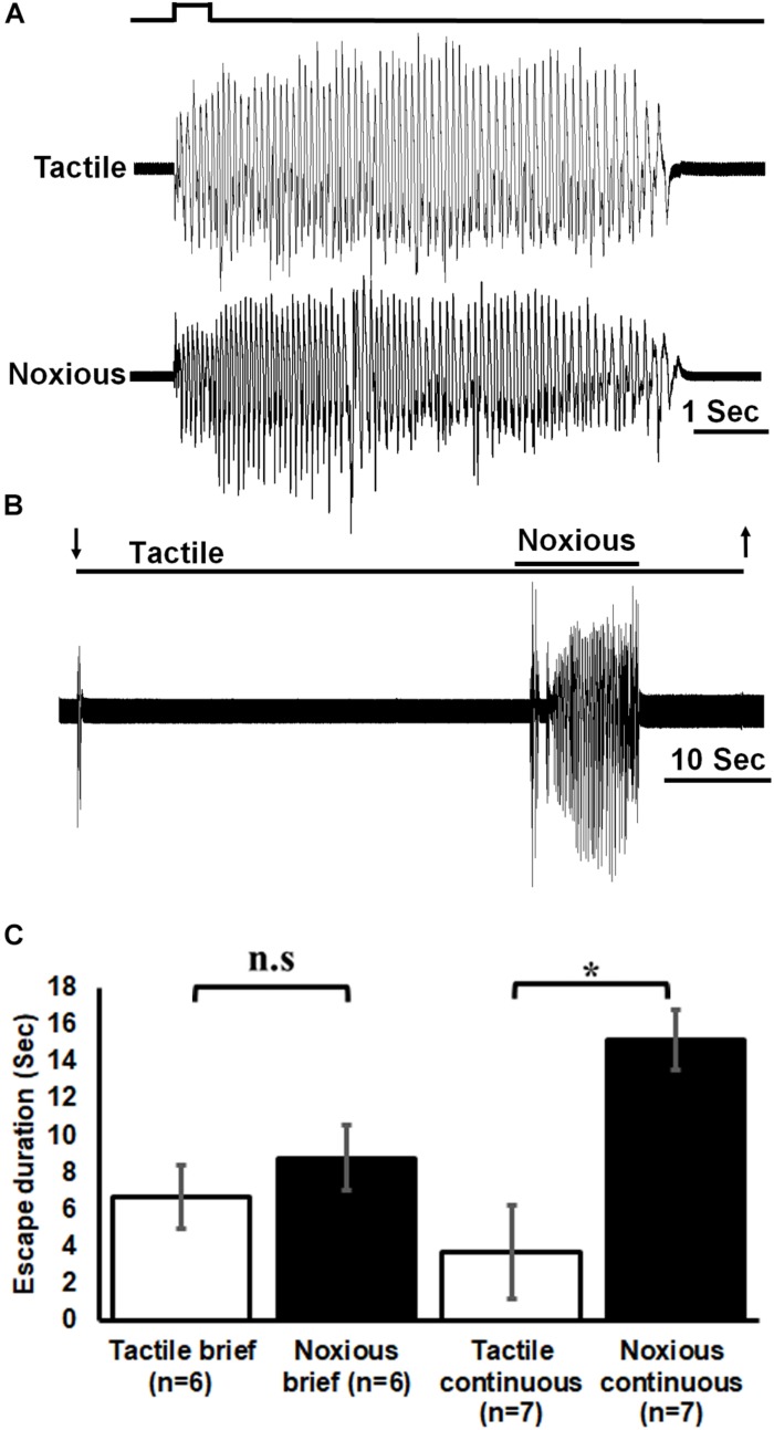
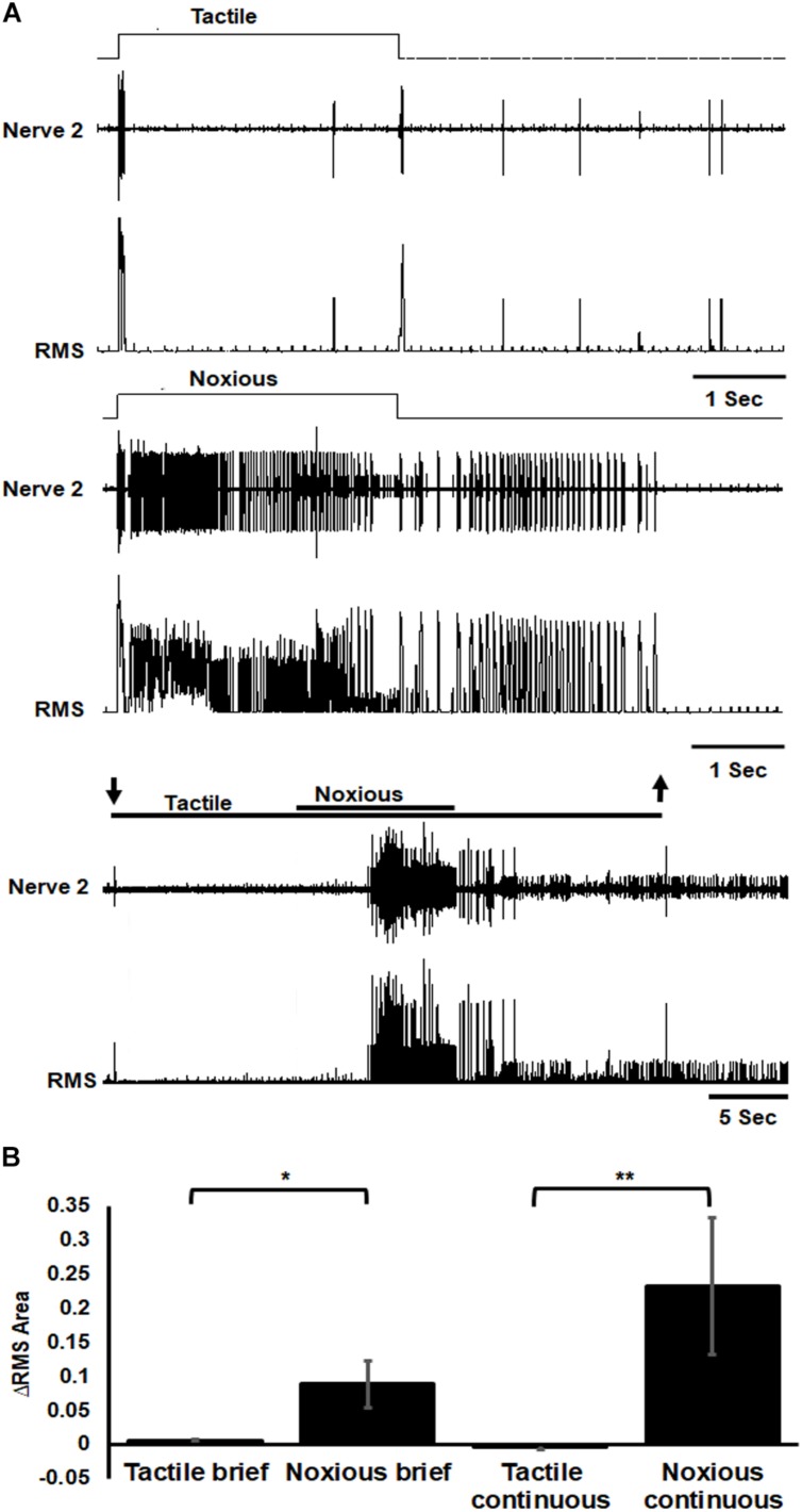
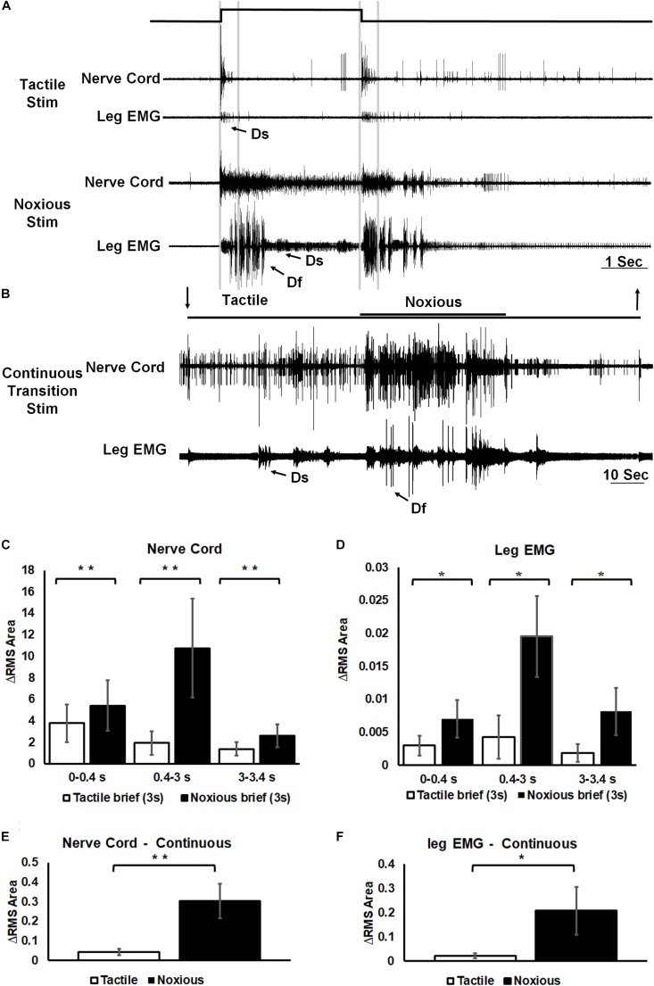
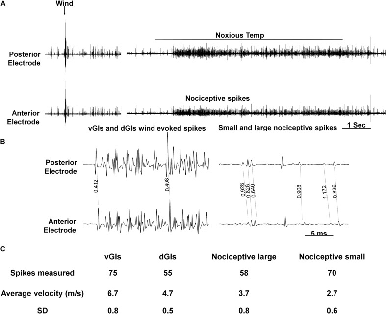
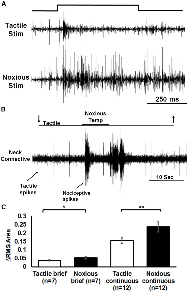
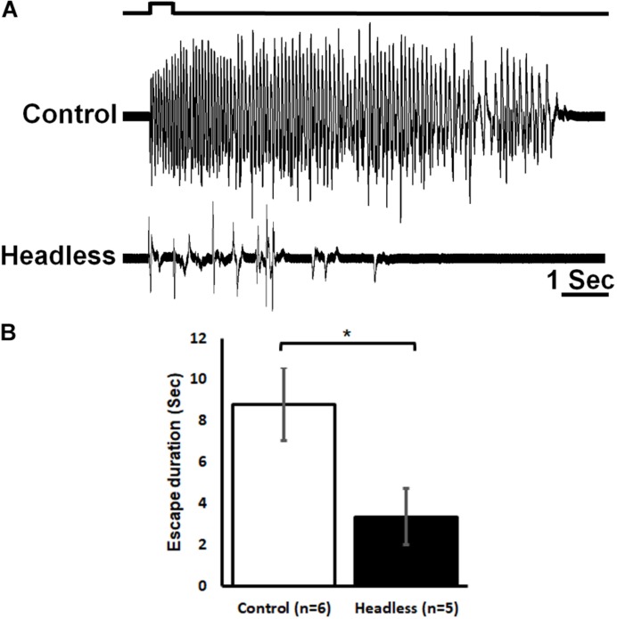
Detecting and avoiding environmental threats such as those with a potential for injury is of crucial importance for an animal's survival. In this work, we examine the nociceptive pathway in an insect, the cockroach Periplaneta americana, from detection of noxious stimuli to nocifensive behavior. We show that noxious stimuli applied to the cuticle of cockroaches evoke responses in sensory axons that are distinct from tactile sensory axons in the sensory afferent nerve. We also reveal differences in the evoked response of post-synaptic projection interneurons in the nerve cord to tactile versus noxious stimuli. Noxious stimuli are encoded in the cockroach nerve cord by fibers of diameter different from that of tactile and wind sensitive fibers with a slower conduction velocity of 2-3 m/s. Furthermore, recording from the neck-connectives show that the nociceptive information reaches the head ganglia. Removing the head ganglia results in a drastic decrease in the nocifensive response indicating that the head ganglia and the nerve cord are both involved in processing noxious stimuli.

Keywords: extracellular recording; insect; interneurons; nociception; nociceptive receptor; nocifensive behavior.


Figures

FIGURE 1
FIGURE 1
A diagram depicting the cockroach’s nervous system. The cockroach’s nervous system comprises two head ganglia (the SupEG or “Brain” and the SEG; black) three thoracic ganglia (T1–T3; dark gray) and six abdominal ganglia (A1–A6; light gray). Two connectives link between adjacent ganglia. The connectives between the head and thorax are termed “neck connectives.” One example of the peripheral Nerve 2 is illustrated on one side of the abdominal ganglion A4. In this preparation Nerve 2 is severed and suction electrode “a” is positioned so the recording is of the sensory input from the cuticle only. Electrodes “b,” “c,” and “d” are shown in the appropriate location along the nervous system and are placed on a single connective.
FIGURE 2
FIGURE 2
Noxious stimulus induces a nocifensive escape response. (A) Leg movements (which represent escape) of a cockroach in response to brief (0.5 s) tactile (upper trace) and noxious (lower trace) stimuli to the abdomen. Stimulus duration is indicated. (B) Leg movements in response to continuous tactile and noxious stimuli to the abdomen. Stimulus duration is indicated with horizontal lines and arrows (upper line: noxious; lower line: tactile). (C) Brief noxious and tactile stimuli display similar escape response. Continuous noxious stimuli induces a significantly higher escape duration than continuous tactile stimuli. Bars represent means ± SEM, significance is indicated with asterisk or with n.s.
FIGURE 3
FIGURE 3
Sensory axons response to noxious stimulus is stronger than the response to tactile stimulus. (A) Extracellular recording of abdominal nerve 2 shows that the response to brief (3 s) tactile stimulus (two upper traces) is transient in the onset and offset of the stimulus while the response to brief (3 s) noxious stimulus (two middle traces) is prolonged and involves different units from that of tactile stimulus. Continuous transition from tactile to noxious stimuli (two lower traces) shows a transient response to tactile stimulus and an ongoing response to noxious stimulus. Stimulus duration is indicated with horizontal lines and arrows (upper line: noxious; lower line: tactile). The upper trace of each stimulus example (tactile, noxious or continuous transition) is the response of the nerve and the lower trace is the same data after RMS procedure. (B) The averaged RMS area shows that the response to brief or continuous noxious stimuli is stronger than that of tactile stimuli. Bars represent means ± SEM, significance is indicated with asterisk.
FIGURE 4
FIGURE 4
Noxious information is carried along the nerve cord and induces escape behavior. (A) Representative example of a simultaneous recordings from post-synaptic interneurons in the nerve cord (upper trace of each stimulus example) and the leg EMG (lower trace of each stimulus example) following brief (3 s) tactile (upper traces) and noxious (lower traces) stimuli. Vertical lines represent a time division (0–0.4, 0.4–3, and 3–3.4 s) corresponding to onset, duration and offset of the stimulus. Activity of the fast and slow motor neurons (Df and Ds, respectively) is indicated on the leg EMG traces. (B) Representative example of a simultaneous recordings from post-synaptic interneurons in the nerve cord (upper trace) and the leg EMG (lower trace) following a continuous transition between tactile and noxious stimuli. Stimulus duration is indicated with horizontal lines and arrows (upper line: noxious; lower line: tactile). Activity of Df and Ds is indicated on the leg EMG trace. (C) Averaged RMS area of nerve cord response to brief tactile and noxious stimuli, according to the mentioned time division. Panel (D) same as panel (B) for the leg EMG response. (E) Averaged RMS area of nerve cord response to a continuous transition between tactile and noxious stimuli. Panel (F) same as panel (E) for the leg EMG response. For panels (C–F) bars represent means ± SEM, significance is indicated with asterisk.
FIGURE 5
FIGURE 5
Conduction velocity of nociceptive projections neurons is two to three-fold slower than that of wind projection interneurons. (A) Response of interneurons in the nerve cord to a wind stimulus (left) and a continuous transition between tactile and noxious stimuli (right). Two electrodes were used to measure velocity (upper and lower traces represent posterior and anterior electrodes, respectively). (B) Enlarged time scale of the traces in panel (A). Time differences (in seconds) between two similar spikes are shown. (C) A table summarizing the data from conduction velocity tests.
FIGURE 6
FIGURE 6
Extracellular recording of neck connective ascending activity following tactile and noxious stimuli shows that noxious information is carried along the nerve cord to the head ganglia. (A) Representative example of neck connective response to brief (0.5 s) tactile (upper trace) or noxious (lower trace) stimuli (stimulus duration is indicated). (B) Activity in the neck connective in response to a continuous transition between tactile and noxious stimuli. Stimulus duration is indicated with horizontal lines and arrows (upper line: noxious; lower line: tactile). (C) The averaged RMS area of the neck connective response to brief (0.5 s)/continuous noxious stimuli is significantly higher than that of brief/continuous tactile stimuli. Bars represent means ± SEM, significance is indicated with asterisk.
FIGURE 7
FIGURE 7
The head ganglia are required for proper escape response to noxious stimuli. (A) Response of control (upper trace) and headless (lower trace) tethered cockroaches to brief (0.5 s) noxious stimuli. (B) The escape response to noxious stimuli of headless cockroaches is reduced as compared to control cockroaches. Bars represent means ± SEM, significance is indicated with asterisk.

Similar articles

Cited by

References

    1. Burrell B. D. (2017). Comparative biology of pain: what invertebrates can tell us about how nociception works. J. Neurophysiol. 117 1461–1473. 10.1152/jn.00600.2016 - DOI - PMC - PubMed
    1. Diwaker A. B. (1973). Distribution and Orientation of Campaniform Sensilla on the Abdomen of the Cockroaches Periplaneta Americana and Blaberus Cranifer. Emporia, KS: Emporia State University.
    1. Duve H., Thorpe A. (1988). Mapping of enkephalin-related peptides in the nervous system of the blowfly, Calliphora vomitoria, and their co-localization with cholecystokinin (CCK)-and pancreatic polypeptide (PP)-like peptides. Cell Tissue Res. 251 399–415. 10.1007/bf00215849 - DOI - PubMed
    1. Dyakonova V. (2001). Role of opioid peptides in behavior of invertebrates. J. Evol. Biochem. Physiol. 37 335–347.
    1. Dyakonova V., Schürmann F.-W., Sakharov D. (1999). Effects of serotonergic and opioidergic drugs on escape behaviors and social status of male crickets. Naturwissenschaften 86 435–437. 10.1007/s001140050647 - DOI - PubMed

LinkOut - more resources