

Emerging Roles of Complement in Psychiatric Disorders
- PMID: 31496960
- PMCID: PMC6712161
- DOI: 10.3389/fpsyt.2019.00573
Emerging Roles of Complement in Psychiatric Disorders
Abstract
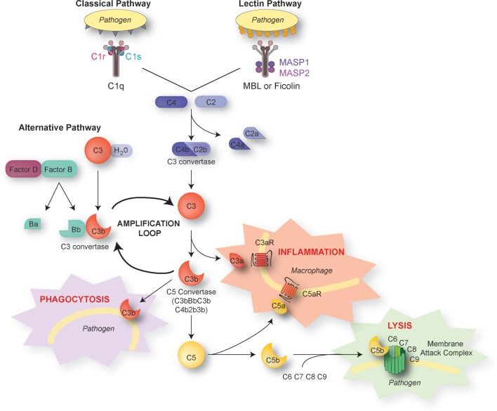
The complement system consists of more than 30 proteins that have long been known to participate to the immune defence against pathogens and to the removal of damaged cells. Their role, however, extends beyond immunity and clearance of altered "self" components in the periphery. In particular, complement proteins can be induced by all cell types in the brain. Recent discoveries highlight the role of complement in normal and pathological brain development. Specifically, the complement system mediates synaptic pruning, a developmental process whereby supernumerary synapses are eliminated in the immature brain. The complement system has been implicated in pathological synapse elimination in schizophrenia, West Nile virus infection, and lupus, all of which are associated with psychiatric manifestations. Complement also contributes to synapse loss in neurodegenerative conditions. This review provides a brief overview of the well-studied role of complement molecules in immunity. The contribution of complement to embryonic and adult neurogenesis, neuronal migration, and developmental synaptic elimination in the normal brain is reviewed. We discuss the role of complement in synapse loss in psychiatric and neurological diseases and evaluate the therapeutic potential of complement-targeting drugs for brain disorders.
Keywords: brain development; microglia; schizophrenia; synapse elimination; synaptic pruning.
Figures

Similar articles
-
Complement System in Neural Synapse Elimination in Development and Disease.Adv Immunol. 2017;135:53-79. doi: 10.1016/bs.ai.2017.06.004. Epub 2017 Jul 31. Adv Immunol. 2017. PMID: 28826529 Review.
-
Complement and microglia dependent synapse elimination in brain development.WIREs Mech Dis. 2022 May;14(3):e1545. doi: 10.1002/wsbm.1545. Epub 2021 Nov 4. WIREs Mech Dis. 2022. PMID: 34738335 Free PMC article. Review.
-
Insight into the role of phosphatidylserine in complement-mediated synapse loss in Alzheimer's disease.Fac Rev. 2021 Feb 24;10:19. doi: 10.12703/r/10-19. eCollection 2021. Fac Rev. 2021. PMID: 33718936 Free PMC article. Review.
-
The complement system: an unexpected role in synaptic pruning during development and disease.Annu Rev Neurosci. 2012;35:369-89. doi: 10.1146/annurev-neuro-061010-113810. Annu Rev Neurosci. 2012. PMID: 22715882 Review.
-
Synaptic Elimination in Neurological Disorders.Curr Neuropharmacol. 2019;17(11):1071-1095. doi: 10.2174/1570159X17666190603170511. Curr Neuropharmacol. 2019. PMID: 31161981 Free PMC article. Review.
Cited by
-
Viral respiratory infections and psychosis: A review of the literature and the implications of COVID-19.Neurosci Biobehav Rev. 2021 Aug;127:520-530. doi: 10.1016/j.neubiorev.2021.05.008. Epub 2021 May 13. Neurosci Biobehav Rev. 2021. PMID: 33992695 Free PMC article. Review.
-
Brain-derived neurotrophic factor from microglia regulates neuronal development in the medial prefrontal cortex and its associated social behavior.Mol Psychiatry. 2024 May;29(5):1338-1349. doi: 10.1038/s41380-024-02413-y. Epub 2024 Jan 19. Mol Psychiatry. 2024. PMID: 38243072 Free PMC article.
-
DLK signaling in axotomized neurons triggers complement activation and loss of upstream synapses.Cell Rep. 2024 Feb 27;43(2):113801. doi: 10.1016/j.celrep.2024.113801. Epub 2024 Feb 14. Cell Rep. 2024. PMID: 38363678 Free PMC article.
-
Regulation of microglia phagocytosis and potential involvement of exercise.Front Cell Neurosci. 2022 Jul 25;16:953534. doi: 10.3389/fncel.2022.953534. eCollection 2022. Front Cell Neurosci. 2022. PMID: 35959472 Free PMC article. Review.
-
Investigating the causal relationships between attention-deficit/hyperactivity disorder and autoimmune diseases: Evidence from Mendelian randomization study.Medicine (Baltimore). 2025 Jan 3;104(1):e41157. doi: 10.1097/MD.0000000000041157. Medicine (Baltimore). 2025. PMID: 40184135 Free PMC article.
References
Publication types
LinkOut - more resources
Full Text Sources