Human T Cell Differentiation Negatively Regulates Telomerase Expression Resulting in Reduced Activation-Induced Proliferation and Survival
- PMID: 31497023
- PMCID: PMC6712505
- DOI: 10.3389/fimmu.2019.01993
Human T Cell Differentiation Negatively Regulates Telomerase Expression Resulting in Reduced Activation-Induced Proliferation and Survival
Abstract
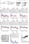
Maintenance of telomeres is essential for preserving T cell proliferative responses yet the precise role of telomerase in human T cell differentiation, function, and aging is not fully understood. Here we analyzed human telomerase reverse transcriptase (hTERT) expression and telomerase activity in six T cell subsets from 111 human adults and found that levels of hTERT mRNA and telomerase activity had an ordered decrease from naïve (TN) to central memory (TCM) to effector memory (TEM) cells and were higher in CD4+ than their corresponding CD8+ subsets. This differentiation-related reduction of hTERT mRNA and telomerase activity was preserved after activation. Furthermore, the levels of hTERT mRNA and telomerase activity were positively correlated with the degree of activation-induced proliferation and survival of T cells in vitro. Partial knockdown of hTERT by an anti-sense oligo in naïve CD4+ cells led to a modest but significant reduction of cell proliferation. Finally, we found that activation-induced levels of telomerase activity in CD4+ TN and TCM cells were significantly lower in old than in young subjects. These findings reveal that hTERT/telomerase expression progressively declines during T cell differentiation and age-associated reduction of activation-induced expression of hTERT/telomerase mainly affects naïve CD4+ T cells and suggest that enhancing telomerase activity could be a strategy to improve T cell function in the elderly.
Keywords: T cell subsets; T lymphocytes; aging; alternative splicing; differentiation; hTERT; proliferation; telomerase.
Figures


Similar articles
-
[Cisplatin-induced apoptotic endonuclease EndoG inhibits telomerase activity and causes malignant transformation of human CD4+ T lymphocytes].Biomed Khim. 2017 Jan;63(1):13-26. doi: 10.18097/PBMC2017630113. Biomed Khim. 2017. PMID: 28251947 Russian.
-
Human normal T lymphocytes and lymphoid cell lines do express alternative splicing variants of human telomerase reverse transcriptase (hTERT) mRNA.Biochem Biophys Res Commun. 2007 Feb 23;353(4):999-1003. doi: 10.1016/j.bbrc.2006.12.149. Epub 2006 Dec 27. Biochem Biophys Res Commun. 2007. PMID: 17204238
-
Alternative splicing of telomerase catalytic subunit hTERT generated by apoptotic endonuclease EndoG induces human CD4+ T cell death.Eur J Cell Biol. 2017 Oct;96(7):653-664. doi: 10.1016/j.ejcb.2017.08.004. Epub 2017 Sep 1. Eur J Cell Biol. 2017. PMID: 28886883
-
Alternative Splicing of hTERT Pre-mRNA: A Potential Strategy for the Regulation of Telomerase Activity.Int J Mol Sci. 2017 Mar 7;18(3):567. doi: 10.3390/ijms18030567. Int J Mol Sci. 2017. PMID: 28272339 Free PMC article. Review.
-
Alternative Splicing of Human Telomerase Reverse Transcriptase (hTERT) and Its Implications in Physiological and Pathological Processes.Biomedicines. 2021 May 9;9(5):526. doi: 10.3390/biomedicines9050526. Biomedicines. 2021. PMID: 34065134 Free PMC article. Review.
Cited by
-
TRF2 inhibition rather than telomerase disruption drives CD4T cell dysfunction during chronic viral infection.J Cell Sci. 2022 Jul 1;135(13):jcs259481. doi: 10.1242/jcs.259481. Epub 2022 Jul 4. J Cell Sci. 2022. PMID: 35660868 Free PMC article.
-
CD8+ T Cell Senescence: Lights and Shadows in Viral Infections, Autoimmune Disorders and Cancer.Int J Mol Sci. 2022 Mar 21;23(6):3374. doi: 10.3390/ijms23063374. Int J Mol Sci. 2022. PMID: 35328795 Free PMC article. Review.
-
Long-term exposure to "low-dose" bisphenol A decreases mitochondrial DNA copy number, and accelerates telomere shortening in human CD8 + T cells.Sci Rep. 2020 Sep 25;10(1):15786. doi: 10.1038/s41598-020-72546-x. Sci Rep. 2020. PMID: 32978426 Free PMC article.
-
Immunosenescence: Aging and Immune System Decline.Vaccines (Basel). 2024 Nov 23;12(12):1314. doi: 10.3390/vaccines12121314. Vaccines (Basel). 2024. PMID: 39771976 Free PMC article. Review.
-
Lipid Microbubble-Conjugated Anti-CD3 and Anti-CD28 Antibodies (Microbubble-Based Human T Cell Activator) Offer Superior Long-Term Expansion of Human Naive T Cells In Vitro.Immunohorizons. 2020 Aug 7;4(8):475-484. doi: 10.4049/immunohorizons.2000056. Immunohorizons. 2020. PMID: 32769179 Free PMC article.
References
Publication types
MeSH terms
Substances
LinkOut - more resources
Full Text Sources
Research Materials

