. 2019 Aug 15;11(8):4946-4956.
eCollection 2019.

The inhibition of SGK1 suppresses epithelial-mesenchymal transition and promotes renal tubular epithelial cell autophagy in diabetic nephropathy

Affiliations

The inhibition of SGK1 suppresses epithelial-mesenchymal transition and promotes renal tubular epithelial cell autophagy in diabetic nephropathy

Langen Zhuang et al. Am J Transl Res. .

Abstract

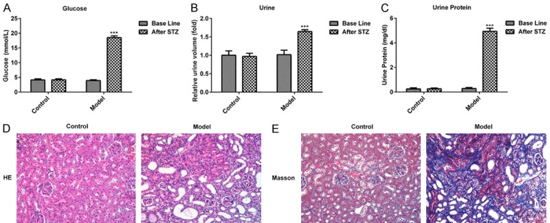
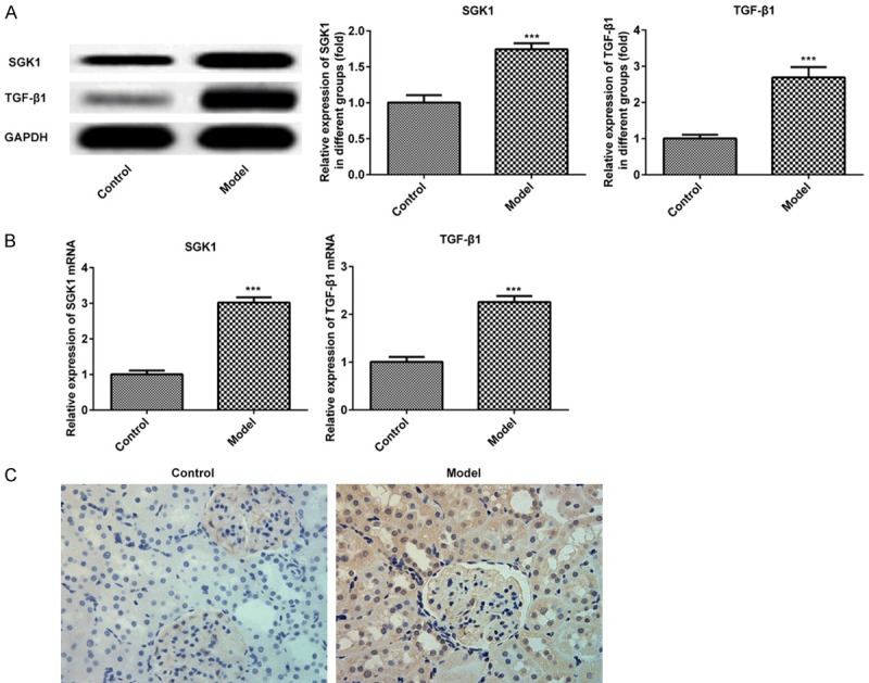
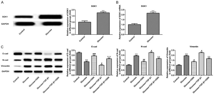
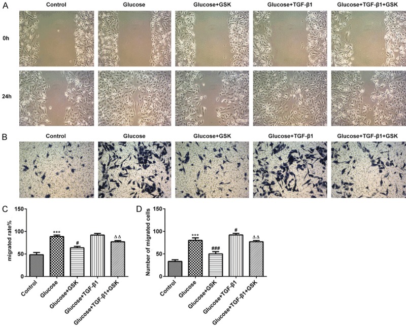
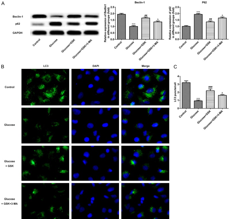
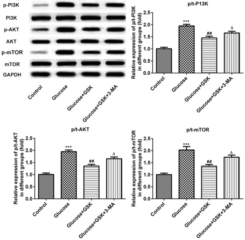
Diabetic nephropathy (DN) is a common complication of diabetes that is the dominant cause of end-stage renal disease. However, the pathological mechanism of DN is yet to be elucidated. Serum and glucocorticoid induced kinase (SGK) 1, a ubiquitously expressed kinase, was employed in the current study to assess its effect on DN in vivo and in vitro. Male BALB/C mice and a human tubular epithelial cell line (HK-2) were utilized for experimentation. Male BALB/C mice and a human tubular epithelial cell line (HK-2) were utilized for experimentation. Pathological changes were measured via HE and staining and immunohistochemistry was performed to measure the expression of SGK 1. An SGK1 inhibitor, GSK650394, was applied to analyze the role of SGK1 in HK-2 cell epithelial-mesenchymal transition (EMT). Associated protein expressions were assessed via western blotting. In addition, migration was measured using a scratch wound healing assay. 3-methyladenine (3-MA), an autophagy inhibitor, was used to determine the variation of autophagy following SGK1 inhibition. The expression of autophagy proteins were analyzed. Furthermore, the expression of PI3K, AKT, mTOR and their levels of phosphorylation were measured. The results revealed that the ultrastructure of renal tissue suffered damage and that the expression of SGK1 was markedly increased. After SGK1 inhibition, HK-2 cell EMT was suppressed and cell migration was attenuated. Furthermore, the autophagy of HK-2 cells was promoted, an increased expression of Beclin-1 and LC3 II was detected, and a decreased expression of p62 was observed. Additionally, the phosphorylation of PI3K, AKT and mTOR were markedly upregulated. The results indicated that blocking autophagy signaling via 3-MA muted SGK1-protected against HG-evoked cell injury. Our study demonstrated that SGK1 inhibition promoted autophagy and suppressed renal tubular epithelial cell EMT in DN, indicating that SGK1 may serve as a potential therapeutic target of DN.

Keywords: Autophagy; diabetic nephropathy; epithelial mesenchymal transition; serum-and glucocorticoid induced kinase.


Conflict of interest statement

None.

Figures

Figure 1
Figure 1
Physiological changes of mice after injection with STZ for 72 h. (A) The levels of glucose, (B) urine volume and (C) urine protein in control and STZ treated mice are presented. The pathologic changes of renal tissues were measured via (D) hematoxylin and eosin and (E) Masson staining. (magnification, ×100). ***P < 0.001 vs. the control. STZ, streptozotocin.
Figure 2
Figure 2
Expression of SGK1 and TGF-β1 in murine renal tissue following treatment with STZ. The expression of SGK1 and TGF-β1 were assessed via (A) western blotting and (B) reverse transcription-quantitative PCR. (C) The expression of SGK1 was determined via immunohistochemistry. (magnification, ×200). ***P < 0.001 vs. the control. SGK1, serum and glucocorticoid induced kinase; TGF-β1, transforming growth factor; STZ, streptozotocin.
Figure 3
Figure 3
Expression of SGK1, E-cadherin, N-cadherin and Vimentin following the treatment of high glucose, TGF-β1 or GSK in HK-2 cells. The expression of SGK1 was measured via (A) western blotting and (B) reverse transcription-quantitative PCR. (C) The expression of E-cadherin, N-cadherin and Vimentin was determined via western blotting. ***P < 0.001 vs. the control; #P < 0.05, ##P < 0.01 and ###P < 0.001 vs. the Glucose group; ΔΔP < 0.01 and ΔΔΔP < 0.001 vs. the Glucose+TGF-β1 group. SGK1, serum and glucocorticoid induced kinase; TGF-β1, transforming growth factor; GSK, GSK650394.
Figure 4
Figure 4
HK-2 cell migration following treatment with high glucose, TGF-β1 or GSK. The migratory activity of HK-2 cells was assessed via (A and C) scratch and (B and D) Transwell assays (magnification, ×20). ***P < 0.001 vs. the control; #P < 0.05 and ###P < 0.001 vs. the Glucose group; ΔΔP < 0.01 vs. the Glucose+TGF-β1 group. TGF-β1, transforming growth factor; GSK, GSK650394.
Figure 5
Figure 5
Expression of Beclin-1, p62 and LC3 following HK-2 cell treatment with high glucose, GSK or 3-MA. A. The expressions of Beclin-1 and p62 were measured via western blotting. B. Levels of LC3 were determined via immunofluorescence. (magnification, ×200). C. Quantitative analysis for immunofluorescence. ***P < 0.001 vs. the control; ##P < 0.01 and ###P < 0.001 vs. the Glucose group; ΔP < 0.05 vs. the Glucose+TGF-β1 group. GSK, GSK650394’ 3-MA, 3-methyladenine.
Figure 6
Figure 6
Expression of p-PI3K, p-AKT and p-mTOR following HK-2 cell treatment with high glucose, GSK or 3-MA. The expressions of p-PI3K, p-AKT and p-mTOR were measured via western blotting. ***P < 0.001 vs. the control; ##P < 0.01 vs. the Glucose group; ΔP < 0.05 vs. the Glucose+TGF-β1 group. p, phosphorylated; GSK, GSK650394’ 3-MA, 3-methyladenine.

References

    1. Reidy K, Kang HM, Hostetter T, Susztak K. Molecular mechanisms of diabetic kidney disease. J Clin Invest. 2014;124:2333–2340. - PMC - PubMed
    1. Badal SS, Danesh FR. New insights into molecular mechanisms of diabetic kidney disease. Am J Kidney Dis. 2014;63:S63–83. - PMC - PubMed
    1. Magee C, Grieve DJ, Watson CJ, Brazil DP. Diabetic nephropathy: a tangled web to unweave. Cardiovasc Drugs Ther. 2017;31:579–592. - PMC - PubMed
    1. Han Q, Zhu H, Chen X, Liu Z. Non-genetic mechanisms of diabetic nephropathy. Front Med. 2017;11:319–332. - PubMed
    1. Palsamy P, Subramanian S. Resveratrol protects diabetic kidney by attenuating hyperglycemia-mediated oxidative stress and renal inflammatory cytokines via Nrf2-Keap1 signaling. Biochim Biophys Acta. 2011;1812:719–731. - PubMed

LinkOut - more resources