Case Reports
. 2019 Jun 25;11(6):e4998.
doi: 10.7759/cureus.4998.

Melanoacanthoma Masquerading as Melanoma: Case Reports and Literature Review

Affiliations
Case Reports

Melanoacanthoma Masquerading as Melanoma: Case Reports and Literature Review

Nikolas Gutierrez et al. Cureus. .

Abstract

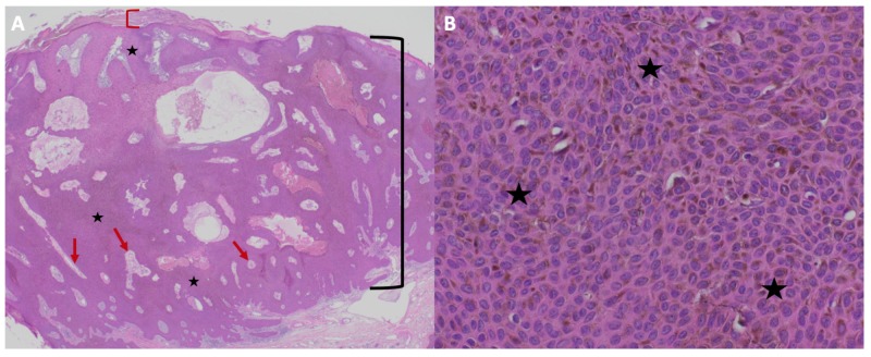
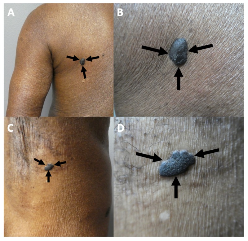
Melanoacanthoma is a benign epithelial tumor composed of melanocytes and keratinocytes that can morphologically mimic malignant neoplasms. Two patients with melanoacanthoma that clinically masqueraded as melanoma are described: a 65-year-old African-American woman with a pigmented nodule on the right preauricular area and an 85-year-old Haitian-Creole man with a large exophytic nodule on his left lower abdomen. Melanoma was clinically suspected in both patients. Biopsies were performed, which established the diagnosis of melanoacanthoma. Complete removal of a melanoacanthoma should be considered since partial excision may result in recurrence.

Keywords: malignant; melanoacanthoma; melanoma; pigmented melanocyte.


Conflict of interest statement

The authors have declared that no competing interests exist.

Figures

Figure 1
Figure 1. Clinical presentation of melanoacanthoma on the right preauricular area of a 65-year-old woman
Clinical presentation of the initial (A) and recurrent (B) melanoacanthoma (black arrows) on the right preauricular area. The tumor initially presented as a 2 x 1-cm black plaque (A). The lesion not only persisted but also increased in size, morphologically mimicking a melanoma (B).
Figure 2
Figure 2. Microscopic presentation of melanoacanthoma on the right preauricular area of a 65-year-old woman
Distant (A) and closer (B-D) views of pathologic features of a melanoacanthoma. Low (A) and higher (B and C) magnification of hematoxylin and eosin stained sections shows an exophytic nodule with hyperkeratosis (thickening of the stratum corneum as shown between the red bracket) and acanthosis (thickening of the epidermis as shown between the black bracket); the area enclosed in the black circle of image A is shown at higher magnification in image B. There is hyperpigmentation throughout all layers of the epidermis (black stars). Tangential sectioning of the tumor shows small areas of dermis, containing epithelial lined vessels and erythrocytes within the epithelium (red arrows). There is lymphocytic perivascular inflammation in the dermis. A higher magnification view (D) of MART-1 stained section shows positive staining of dendritic melanocytes throughout all layers of the epidermis (black arrows) (Hematoxylin and eosin: A, x2; B, x10; C, x40; MART-1 immunoperoxidase; D, x40).
Figure 3
Figure 3. Microscopic presentation of recurrent melanoacanthoma on the right preauricular area of a 65-year-old woman
Distant (A) and closer (B) views of pathologic features of a melanoacanthoma. Low (A) and higher (B) magnification of hematoxylin and eosin stained sections show an exophytic nodule with hyperkeratosis (thickening of the stratum corneum as shown between the red bracket) and acanthosis (thickening of the epidermis as shown between the black bracket). There is hyperpigmentation throughout all layers of the epidermis (black stars). Tangential sectioning of the tumor shows small areas of dermis, containing epithelial lined vessels and erythrocytes within the epithelium (red arrows). There is lymphocytic perivascular inflammation in the dermis (Hematoxylin and eosin: A, x2; B, x40).
Figure 4
Figure 4. Melanoacanthoma on the left lower abdomen of an 85-year-old man
Distant (A) and closer (B) views of a melanoacanthoma (black arrows) presenting as a large 3 x 2.5-cm lobulated, exophytic, black nodule with hyperpigmentation of the surrounding skin on the left lower abdomen.
Figure 5
Figure 5. Pigmented seborrheic keratoses on the left posterior shoulder and the left axilla on an 85-year-old man.
The left posterior shoulder (A and B) and left axilla (C and D) show distant (A and C) and closer (B and D) views of pigments seborrheic keratoses (black arrows) in a man with concurrent melanoacanthoma on his left lower abdomen.
Figure 6
Figure 6. Microscopic presentation of melanoacanthoma on the left lower abdomen of an 85-year-old man
The melanoacanthoma on the lower abdomen of an 85-year-old man contains exophytic (A and B) and endophytic (C and D) portions. Low (A) and higher (B) magnification of the exophytic portion shows hyperkeratosis (thickening of the stratum corneum as shown between the red bracket) and acanthosis (thickening of the epidermis as shown between the black bracket); the area enclosed in the black circle of image A is shown at higher magnification in image B. There is hyperpigmentation throughout all layers of the epidermis (black stars). Tangential sectioning of the tumor shows not only keratin-filled pseudocysts (yellow arrows) but also small areas of dermis containing epithelial lined vessels and erythrocytes within the epithelium (red arrows). There is lymphocytic perivascular inflammation in the dermis. Low (C) and higher (D) magnification of the endophytic portion shows similar pathologic changes; the area enclosed in the black circle of image C is shown at higher magnification in image D. In addition, the higher magnification view (D) shows dendritic melanocytes throughout all layers of the epidermis (black arrows) and hyperpigmentation throughout all layers of the epidermis (black stars; Hematoxylin and eosin: A, x2; B, x10; C, x40; D, x40).
Figure 7
Figure 7. Microscopic presentation of pigmented seborrheic keratoses on the left posterior shoulder and left axilla of an 85-year-old man with concurrent melanoacanthoma on his left lower abdomen.
The left posterior shoulder (A) and left axilla (B) show complete views of pigmented seborrheic keratoses in an 85-year-old man. Pathologic features include hyperkeratosis (thickening of the stratum corneum as shown between the red bracket), acanthosis (thickening of the epidermis as shown between the black bracket), and hyperpigmentation restricted to the basal layers of the epidermis (black stars). Tangential sectioning of the tumor also shows only keratin-filled pseudocysts (yellow arrows) (Hematoxylin and eosin: A, x2; B, x2).

Similar articles

Cited by

References

    1. Clinical and dermoscopic features of cutaneous melanoacanthoma. Chung E, Marghoob A, Carrera C, et al. JAMA Dermatol. 2015;151:1129–1130. - PubMed
    1. Benign mixed tumors of melanocytes and malpighian cells. Mishima Y, Pinkus H. AMA Arch Dermatol. 1960;81:539–550. - PubMed
    1. Melanoacanthoma: ultrastructural and immunological studies. Schlappner OL, Rowden G, Philips TM, Rahim Z. J Cutan Pathol. 1978;5:127–141. - PubMed
    1. Giant melanoacanthoma mimicking malignant melanoma. Shankar V, Nandi J, Ghosh K, Ghosh S. Indian J Dermatol. 2011;56:79–81. - PMC - PubMed
    1. Melanoacanthoma: uncommon presentation of an uncommon condition. Vasani RJ, Khatu SS. Indian Dermatol Online J. 2013;4:119–121. - PMC - PubMed

Publication types

LinkOut - more resources