

Case Reports
doi: 10.7759/cureus.5010.
Brain Metastasis in a Young Patient with Uterine Carcinosarcoma
Affiliations
- PMID: 31497440
- PMCID: PMC6713238
- DOI: 10.7759/cureus.5010
Item in Clipboard
Case Reports
Brain Metastasis in a Young Patient with Uterine Carcinosarcoma
Cureus.
.
Abstract
Uterine carcinosarcoma occurs almost exclusively in post-menopausal women and often carries high rates of disease recurrence and mortality. Central nervous system involvement is extremely rare with less than 10 reported cases in the literature. Here we present a young pre-menopausal patient with metastatic malignant mixed Mullerian tumor who developed cerebral metastasis while on systemic chemotherapy.
Keywords: cerebral metasatasis; cns metastasis; endometrial cancer; malignant mixed mullerian tumor; mmmt; uterine carcinosarcoma.
Conflict of interest statement
The authors have declared that no competing interests exist.
Figures

Panes A (sagittal) and B (coronal) reveal diffuse distention and heterogeneous attenuation centered within the endometrium with diffuse enlargement (15 x 13.5 x13 cm) of the uterus (red arrows).

Red arrow points to largest metastatic lung nodule located in the left lower lobe and measuring approximately 1.1 cm in the long axis.
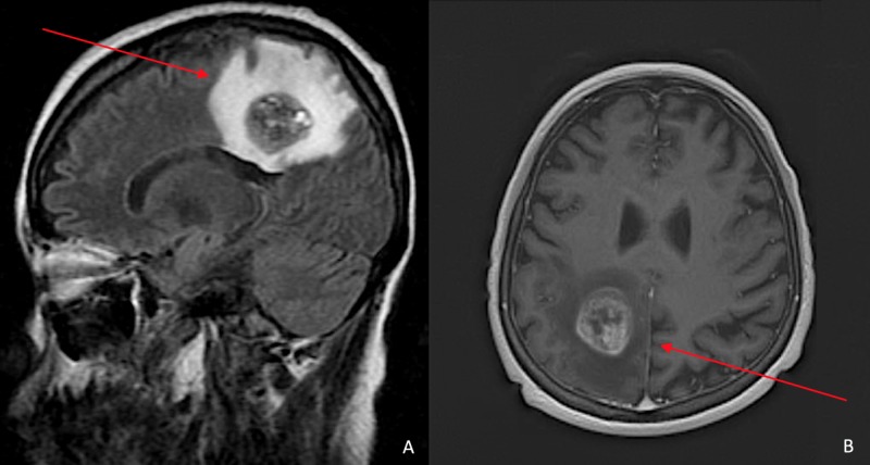
Heterogeneous mass in the parasagittal right parietal lobe just above the posterior body of the corpus callosum (red arrows). It measures 3.0 x 2.7 x 2.5 cm and demonstrates peripheral and septated internal enhancement of IV contrast. There is a large surrounding perilesional vasogenic edema that causes mild mass effect and partial effacement of the posterior horn of the right lateral ventricle with slight inferior displacement of the posterior body of the corpus callosum.
References
-
- Uterine carcinosarcoma: A review of the literature. Cantrell LA, Blank SV, Duska LR. Gynecol Oncol. 2015;137:581–588. - PubMed
-
- Review literature on uterine carcinosarcoma. Singh R. https://www.ncbi.nlm.nih.gov/pubmed/25313723. J Cancer Res Ther. 2014;10:461–468. - PubMed
-
- The significance of epithelial differentiation in mixed mesodermal tumors of the uterus. A clinicopathologic and immunohistochemical study. Bitterman P, Chun B, Kurman R. Am J Surg Pathol. 1990;14:317–328. - PubMed
Publication types
LinkOut - more resources
Full Text Sources