. 2019 Sep;9(9):341.
doi: 10.1007/s13205-019-1874-6. Epub 2019 Aug 23.

Cloning and identification of antimicrobial peptide, hepcidin from freshwater carp, Catla catla on pathogen challenge and PAMPs stimulation

Affiliations

Cloning and identification of antimicrobial peptide, hepcidin from freshwater carp, Catla catla on pathogen challenge and PAMPs stimulation

Rajanya Banerjee et al. 3 Biotech. 2019 Sep.

Abstract

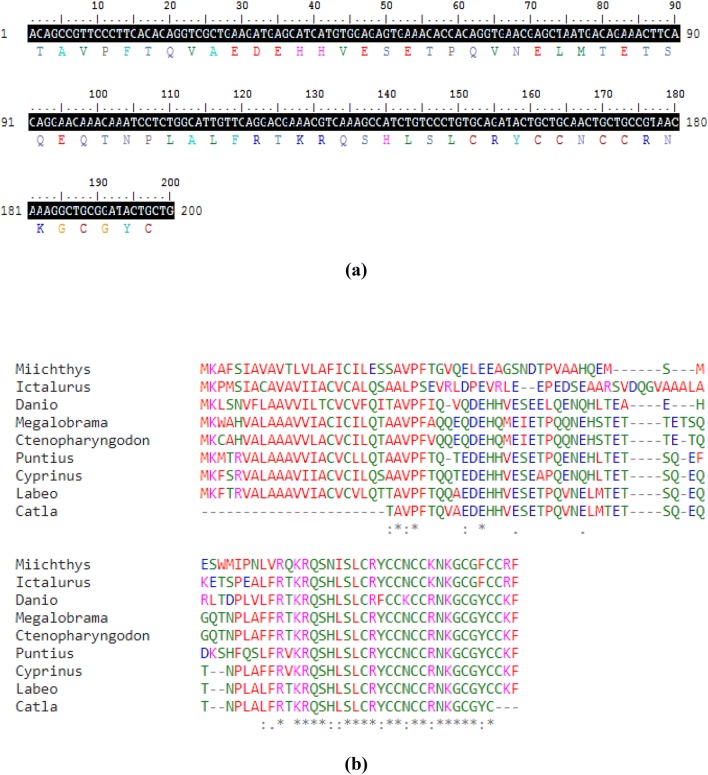
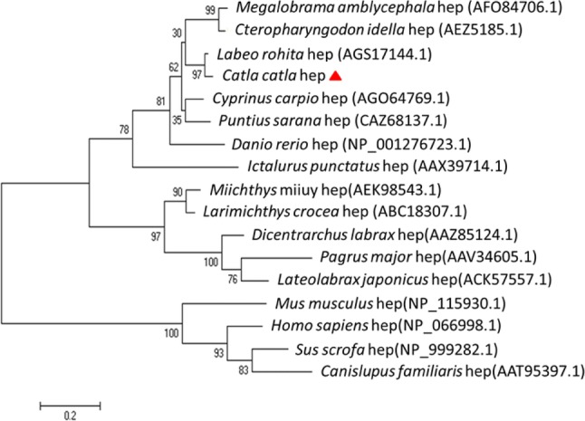
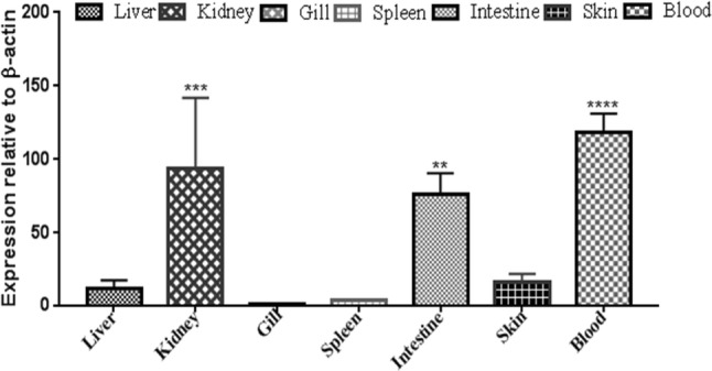
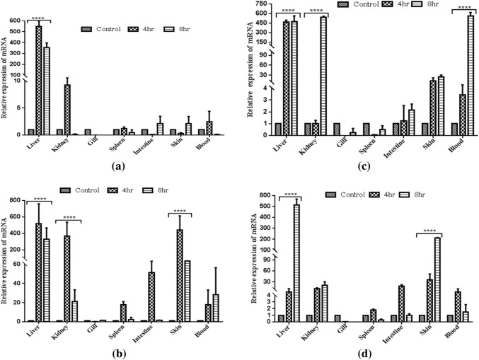
Hepcidin, a cationic cysteine-rich antimicrobial peptide (AMP) acts in hormone regulation and iron homeostasis in the host body. However, the biological property of hepcidin in immune reaction remains unexplored. In aquatic milieu, environmental and pathogenic stressors cause detrimental infections, which are defended by various immunological cells and antimicrobial peptides. In this study, hepcidin gene has been cloned from freshwater carp, Catla catla. The partially cloned hepcidin consists of 200 bp nucleotide sequence encoding 66 amino acids. Nucleotide sequence showed 97% and 91% similarity with Labeo rohita and Cyprinus carpio, respectively. Expression profile revealed significant up-regulation (P ≤ 0.0001) in liver as compared to other tissues in different conditions. In Aeromonas hydrophila challenged C. catla, liver showed higher expression level of hepcidin at 72 h as compared to other tissues. In skin, hepcidin expression showed significant upraise during 24 h in Streptococcus uberis infection. In Argulus sp. infected fishes, up-regulation of hepcidin expression was noted in liver, intestine and skin. The inactivated viral antigen-stimulated fishes, a substantial rise in liver was observed implying hepcidin as an important molecule in combating the pathogenic infections in freshwater carp, C. catla. Fishes stimulated with pathogen-associated molecular patterns (PAMPs) triggered the increased expression of hepcidin mRNA in liver, kidney and skin. This study indicates the presence of hepcidin as antimicrobial peptide in neutralizing the pathogenic infection in fishes.

Keywords: Antimicrobial peptide; Catla catla; Hepcidin; PAMPs; Pathogen challenge; Transcriptomic expression.


Conflict of interest statement

Conflict of interestThe authors declare no conflict of interest.

Figures

Fig. 1
Fig. 1
Sequence analysis of cloned Cchepcidin. a Amino acid deduced from the partial nucleotide sequences of Catla catla mRNA transcript. The protein sequence is shown below the nucleotide sequence (shaded). b Multiple protein sequence alignment of Catla catla hepcidin along with other known submitted sequences are shown using Clustal Omega. The asterisks (*) indicate the identical residues and dots and colons indicate the similar amino acids
Fig. 2
Fig. 2
Bioinformatic analysis: SMART analysis of the derived hepcidin protein sequence
Fig. 3
Fig. 3
Unrooted phylogenetic tree showing the functional relationship between hepcidin constant region sequences of different fish species and the protein sequences were aligned using Clustal Omega. The tree was constructed using the neighbor-joining (NJ) method along with 1000 bootstrap replications using MEGA 6 program. The divergence time is indicated with the help of scale bar
Fig. 4
Fig. 4
Basal expression level of Cchepcidin in varied immunologically relevant tissues. The expression level was represented as a ratio relative to β-actin (internal control) level in the sample. The fold change (2−δδCt) was calculated, where Ct (target) – Ct (internal control) = δCt and δδCt = δCt(target) – δCt(calibrator). β-actin was taken as an internal control. Gill tissue is used as a calibrator. The results were expressed as mean ± standard deviation from three samples (n = 3). Significant differences were determined with one-way ANOVA using PRISM 5 software
Fig. 5
Fig. 5
Transcriptomic Cchepcidin expression on bacterial infection in Catla catla. a Aeromonas hydrophila, and b Streptococcus uberis. The immunologically relevant tissues were processed for qRT-PCR analysis after 24 h, 48 h and 72 h of infection. β-actin was used as an endogenous control. Normalized untreated samples were used as a calibrator to evaluate relative Cchepcidin expression in all tissues at various treatment conditions in terms of fold changes. The results were expressed as mean ± standard deviation from three samples (n = 3). Significant differences among treatment conditions were evaluated by two-way ANOVA using PRISM 5 with ***P < 0.001 and **P < 0.01 as significant levels
Fig. 6
Fig. 6
Transcriptomic Cchepcidin expression on Argulus infection in Catla catla. The immunologically relevant tissues were processed for qRT-PCR analysis after moderate and heavy infection. β-actin was used as an endogenous control. Normalized untreated samples were used as a calibrator to evaluate relative Cchepcidin expression in all tissues at various treatment conditions in terms of fold changes. The results were expressed as mean ± standard deviation from three samples (n = 3). Significant differences among treatment conditions were evaluated by two-way ANOVA using PRISM 5 with *P < 0.05 as significant levels
Fig. 7
Fig. 7
Transcriptomic Cchepcidin expression on inactivated rhabdovirus antigenic stimulation in Catla catla. The immunologically relevant tissues were processed for qRT-PCR analysis after 24 h, 48 h and 72 h of infection. β-actin was used as endogenous control. Normalized untreated samples were used as calibrator to evaluate relative Cchepcidin expression in all tissues at various treatment conditions in terms of fold changes. The results were expressed as mean ± standard deviation from three samples (n = 3). Significant differences among treatment conditions were evaluated by two-way ANOVA using PRISM 5 with ***P < 0.001 and **P < 0.01 as significant levels
Fig. 8
Fig. 8
qRT-PCR analysis of Cchepcidin transcript expression after PAMPs stimulation in various tissues. a Peptidoglycan, b polyinosinic: polycytidylic (poly I:C), c lipopolysaccharide, and d flagellin. RNA extraction of the treated tissues was performed after 4 h and 8 h of stimulation and processed for qRT-PCR analysis. β-actin was used as an endogenous control. Normalized untreated samples were used as a calibrator to evaluate relative Cchepcidin expression in all tissues at various treatment conditions in terms of fold changes. The results were expressed as mean ± standard deviation from three samples (n = 3). Two-way ANOVA was used for determination of the significant differences using PRISM 5 with ***P < 0.001, **P < 0.01 and *P < 0.05 as significant levels

References

    1. Adamek M, Syakuri H, Harris S, Rakus KŁ, Brogden G, Matras M, Steinhagen D. Cyprinid herpesvirus 3 infection disrupts the skin barrier of common carp (Cyprinus carpio L.) Vet Microbiol. 2013;162(2−4):456–470. doi: 10.1016/j.vetmic.2012.10.033. - DOI - PubMed
    1. Banerjee R, Patel B, Basu M, Lenka SS, Paicha M, Samanta M, Das S. Molecular cloning, characterization and expression of immunoglobulin D on pathogen challenge and pathogen associated molecular patterns stimulation in freshwater carp, Catla catla. Microbiol Immunol. 2017;61(10):452–458. doi: 10.1111/1348-0421.12534. - DOI - PubMed
    1. Basu M, Swain B, Maiti NK, Routray P, Samanta M. Inductive expression of toll-like receptor 5 (TLR5) and associated downstream signaling molecules following ligand exposure and bacterial infection in the Indian major carp, mrigal (Cirrhinus mrigala) Fish Shellfish Immunol. 2012;32(1):121–131. doi: 10.1016/j.fsi.2011.10.031. - DOI - PubMed
    1. Bode C, Zhao G, Steinhagen F, Kinjo T, Klinman DM. CpG DNA as a vaccine adjuvant. Expert Rev Vaccines. 2011;10(4):499–511. doi: 10.1586/erv.10.174. - DOI - PMC - PubMed
    1. Bury N, Grosell M. Iron acquisition by teleost fish. Comp Biochem Physiol C Toxicol Pharmacol. 2003;135(2):97–105. doi: 10.1016/S1532-0456(03)00021-8. - DOI - PubMed

LinkOut - more resources