

The Mitochondria-Endoplasmic Reticulum Contacts and Their Critical Role in Aging and Age-Associated Diseases
- PMID: 31497601
- PMCID: PMC6712070
- DOI: 10.3389/fcell.2019.00172
The Mitochondria-Endoplasmic Reticulum Contacts and Their Critical Role in Aging and Age-Associated Diseases
Abstract
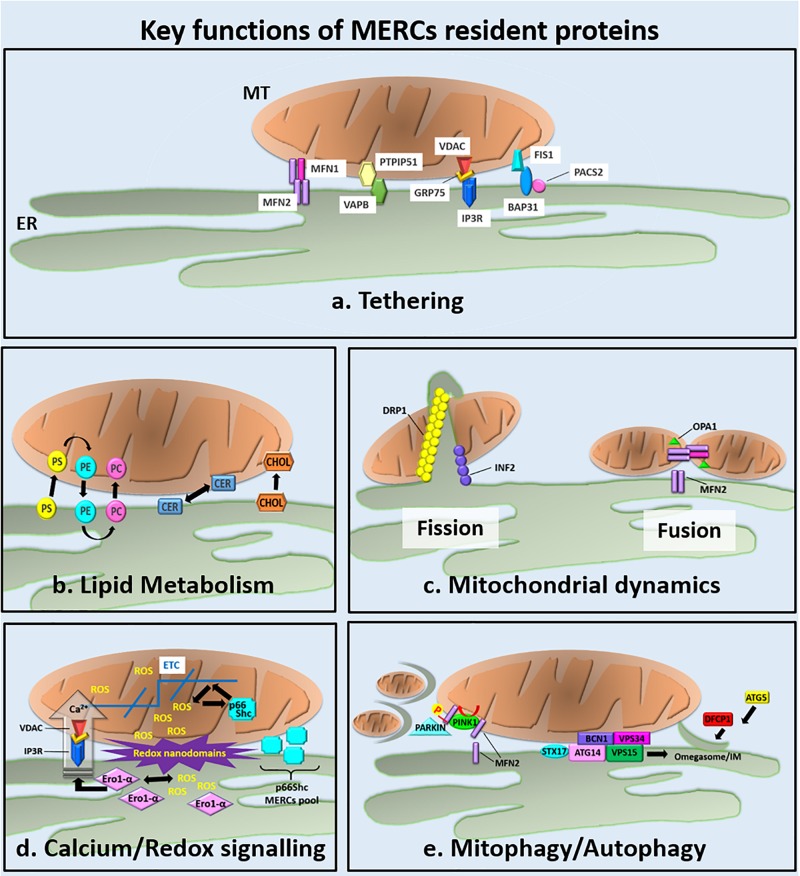
The recent discovery of interconnections between the endoplasmic reticulum (ER) membrane and those of almost all the cell compartments is providing novel perspectives for the understanding of the molecular events underlying cellular mechanisms in both physiological and pathological conditions. In particular, growing evidence strongly supports the idea that the molecular interactions occurring between ER and mitochondrial membranes, referred as the mitochondria (MT)-ER contacts (MERCs), may play a crucial role in aging and in the development of age-associated diseases. As emerged in the last decade, MERCs behave as signaling hubs composed by structural components that act as critical players in different age-associated disorders, such as neurodegenerative diseases and motor disorders, cancer, metabolic syndrome, as well as cardiovascular diseases. Age-associated disorders often derive from mitochondrial or ER dysfunction as consequences of oxidative stress, mitochondrial DNA mutations, accumulation of misfolded proteins, and defective organelle turnover. In this review, we discuss the recent advances associating MERCs to aging in the context of ER-MT crosstalk regulating redox signaling, ER-to MT lipid transfer, mitochondrial dynamics, and autophagy.
Keywords: age-associated diseases; aging; endoplasmic reticulum; mitochondria; oxidative stress; senescence.
Figures
Similar articles
-
Cellular senescence links mitochondria-ER contacts and aging.Commun Biol. 2021 Nov 24;4(1):1323. doi: 10.1038/s42003-021-02840-5. Commun Biol. 2021. PMID: 34819602 Free PMC article. Review.
-
Mitochondria-endoplasmic reticulum contacts in sepsis-induced myocardial dysfunction.Front Cell Dev Biol. 2022 Nov 24;10:1036225. doi: 10.3389/fcell.2022.1036225. eCollection 2022. Front Cell Dev Biol. 2022. PMID: 36506093 Free PMC article. Review.
-
Perspective: Mitochondria-ER Contacts in Metabolic Cellular Stress Assessed by Microscopy.Cells. 2018 Dec 21;8(1):5. doi: 10.3390/cells8010005. Cells. 2018. PMID: 30577576 Free PMC article. Review.
-
Dysfunctional mitochondria as critical players in the inflammation of autoimmune diseases: Potential role in Sjögren's syndrome.Autoimmun Rev. 2021 Aug;20(8):102867. doi: 10.1016/j.autrev.2021.102867. Epub 2021 Jun 9. Autoimmun Rev. 2021. PMID: 34118452 Review.
-
Epigenetic regulation of mitochondrial-endoplasmic reticulum dynamics in kidney diseases.J Cell Physiol. 2023 Aug;238(8):1716-1731. doi: 10.1002/jcp.31058. Epub 2023 Jun 25. J Cell Physiol. 2023. PMID: 37357431 Review.
Cited by
-
Ryanodine Receptors: A Potential Treatment Target in Various Neurodegenerative Disease.Cell Mol Neurobiol. 2021 Nov;41(8):1613-1624. doi: 10.1007/s10571-020-00936-w. Epub 2020 Aug 24. Cell Mol Neurobiol. 2021. PMID: 32833122 Free PMC article. Review.
-
Direct neuronal reprogramming of NDUFS4 patient cells identifies the unfolded protein response as a novel general reprogramming hurdle.Neuron. 2024 Apr 3;112(7):1117-1132.e9. doi: 10.1016/j.neuron.2023.12.020. Epub 2024 Jan 23. Neuron. 2024. PMID: 38266647 Free PMC article.
-
Targeting Mitochondrial Dysfunction and Reactive Oxygen Species for Neurodegenerative Disease Treatment.Int J Mol Sci. 2024 Jul 21;25(14):7952. doi: 10.3390/ijms25147952. Int J Mol Sci. 2024. PMID: 39063194 Free PMC article. Review.
-
Interorganelle communication, aging, and neurodegeneration.Genes Dev. 2021 Apr 1;35(7-8):449-469. doi: 10.1101/gad.346759.120. Genes Dev. 2021. PMID: 33861720 Free PMC article. Review.
-
The Role of Autophagy in the Function of CD4+ T Cells and the Development of Chronic Inflammatory Diseases.Front Pharmacol. 2022 Mar 22;13:860146. doi: 10.3389/fphar.2022.860146. eCollection 2022. Front Pharmacol. 2022. PMID: 35392563 Free PMC article. Review.
References
Publication types
LinkOut - more resources
Full Text Sources
Research Materials