Review
. 2019 Sep 7;394(10201):869-881.
doi: 10.1016/S0140-6736(19)31894-X.

Acute liver failure

Affiliations
Review

Acute liver failure

R Todd Stravitz et al. Lancet. .

Abstract

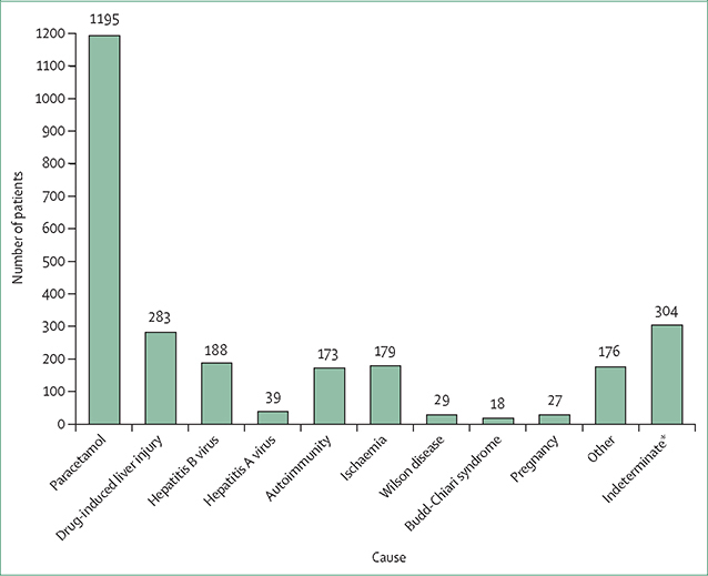
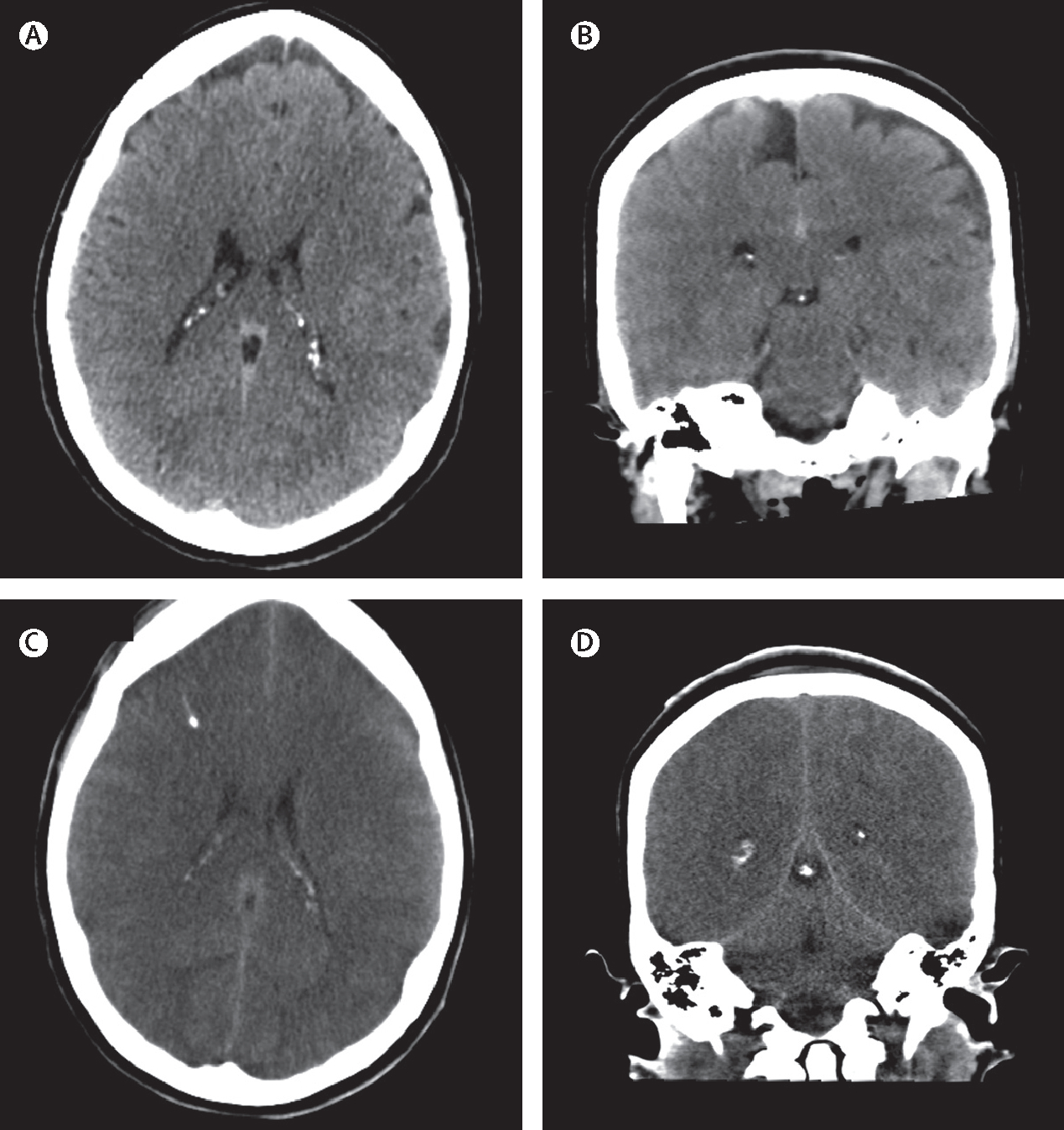
Acute liver failure is a rare and severe consequence of abrupt hepatocyte injury, and can evolve over days or weeks to a lethal outcome. A variety of insults to liver cells result in a consistent pattern of rapid-onset elevation of aminotransferases, altered mentation, and disturbed coagulation. The absence of existing liver disease distinguishes acute liver failure from decompensated cirrhosis or acute-on-chronic liver failure. Causes of acute liver failure include paracetamol toxicity, hepatic ischaemia, viral and autoimmune hepatitis, and drug-induced liver injury from prescription drugs, and herbal and dietary supplements. Diagnosis requires careful review of medications taken, and serological testing for possible viral exposure. Because of its rarity, acute liver failure has not been studied in large, randomised trials, and most treatment recommendations represent expert opinion. Improvements in management have resulted in lower mortality, although liver transplantation, used in nearly 30% of patients with acute liver failure, still provides a life-saving alternative to medical management.


Conflict of interest statement

Declaration of interests

WML has received research grant support from Merck, Conatus, Intercept, Bristol-Myers Squibb, Novo Nordisk, Synlogic, Eiger, Cumberland, Exalenz Bioscience Company, Instrumentation Laboratory, and Ocera Therapeutics (now Mallinckrodt Pharmaceuticals), in the past 36 months. He has received personal fees for consulting from Novartis, Sanofi, and Genentech. RTS has received grant support from Instrumentation Laboratory, Exalenz Bioscience Company, and Ocera Therapeutics (now Mallinckrodt Pharmaceuticals), in the past 36 months.

Figures

Figure 1:
Figure 1:. Causes of acute liver failure, as recorded by the site principal investigator in the US Adult Acute Liver Failure Study Group Registry
Data were collected between Jan 1, 1998 and March 31, 2019. The total number of patients enrolled is 2614; paracetamol accounts for 46% of cases, 12% of cases have indeterminate causes, and drug-induced liver injury accounts for 11% of cases. *The number of patients with acute liver failure with an indeterminate cause decreased to 161, or 5·5% of the total, after review.
Figure 2:
Figure 2:. Examples of histopathology of acute liver failure
(A) Normal liver at low power (100×). (B) Higher power (400×) view of a normal hepatic lobule showing a portal tract. (C) Higher power (400×) view of a normal hepatic lobule showing central hepatic vein. (D) Isoniazid toxicity with complete parenchymal loss, no hepatocytes remain and the normal parenchyma has been replaced by ductular reaction (400×). (E) Heterogeneous areas of necrosis and parenchymal regeneration observed with nitrofurantoin toxicity (40×). (F) Regenerative nodules are rimmed by chronic inflammation at higher power (100×) in the same case as in (E); the patient had taken nitrofurantoin intermittently for nearly 6 months before developing liver failure. Regenerative nodules are often seen in this setting and might be confused on imaging with cirrhosis. (G) Paracetamol toxicity with zone 3 (centrilobular) necrosis. Many hepatocytes show steatosis and in the necrotic zone, lipid drops have coalesced (200×). (H) Acute liver failure related to hepatitis B, with prominent plasma-cell infiltrate within a portal tract, a unique but common finding in fulminant hepatitis B (600×). Representative explants were drawn from the Acute Liver Failure Study Group pathology registry, courtesy of David E Kleiner, Laboratory of Pathology, National Cancer Institute, Bethseda, MD, USA.
Figure 3:
Figure 3:. CT of the head in a patient with acute liver failure who developed cerebral oedema
(A) Axial scan before the development of cerebral oedema. (B) Coronal scan before the development of cerebral oedema. (C) Axial scan after the development of cerebral oedema. (D) Coronal scan after development of cerebral oedema. All in a dying patient with brainstem herniation due to paracetamol-induced acute liver failure. (C) and (D) show the loss of grey–white demarcation and effacement of sulci.

Comment in

  • Acute liver failure.
    Vento S, Cainelli F. Vento S, et al. Lancet. 2020 Jun 13;395(10240):1833. doi: 10.1016/S0140-6736(20)30046-5. Lancet. 2020. PMID: 32534641 No abstract available.

References

    1. Ganger DR, Rule J, Rakela J, et al. Acute liver failure of indeterminate etiology: a comprehensive systematic approach by an expert committee to establish causality. Am J Gastroenterol 2018; 113: 1319–28. - PMC - PubMed
    1. Trey C, Davidson CS. The management of fulminant hepatic failure. Prog Liver Dis 1970; 3: 282–98. - PubMed
    1. Bernal W, Jalan R, Quaglia A, Simpson K, Wendon J, Burroughs A. Acute-on-chronic liver failure. Lancet 2015; 386: 1576–87. - PubMed
    1. Wijdicks EFM. Hepatic encephalopathy. N Engl J Med 2017; 376: 186. - PubMed
    1. Lee WM, Squires RH Jr, Nyberg SL, Doo E, Hoofnagle JH. Acute liver failure: summary of a workshop. Hepatology 2008; 47: 1401–15. - PMC - PubMed