. 2019 Oct 1;76(10):1240-1251.
doi: 10.1001/jamaneurol.2019.2917.

Scientific Advances in and Clinical Approaches to Small-Fiber Polyneuropathy: A Review

Affiliations

Scientific Advances in and Clinical Approaches to Small-Fiber Polyneuropathy: A Review

Anne Louise Oaklander et al. JAMA Neurol. .

Erratum in

  • Error in Affiliation.
    [No authors listed] [No authors listed] JAMA Neurol. 2019 Dec 1;76(12):1520. doi: 10.1001/jamaneurol.2019.3877. JAMA Neurol. 2019. PMID: 31682711 Free PMC article. No abstract available.

Abstract

Importance: Small-fiber polyneuropathy involves preferential damage to the thinly myelinated A-delta fibers, unmyelinated C sensory fibers, or autonomic or trophic fibers. Although this condition is common, most patients still remain undiagnosed and untreated because of lagging medical and public awareness of research advances. Chronic bilateral neuropathic pain, fatigue, and nausea are cardinal symptoms that can cause disability and dependence, including pain medication dependence.

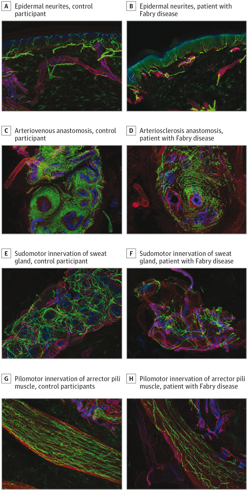
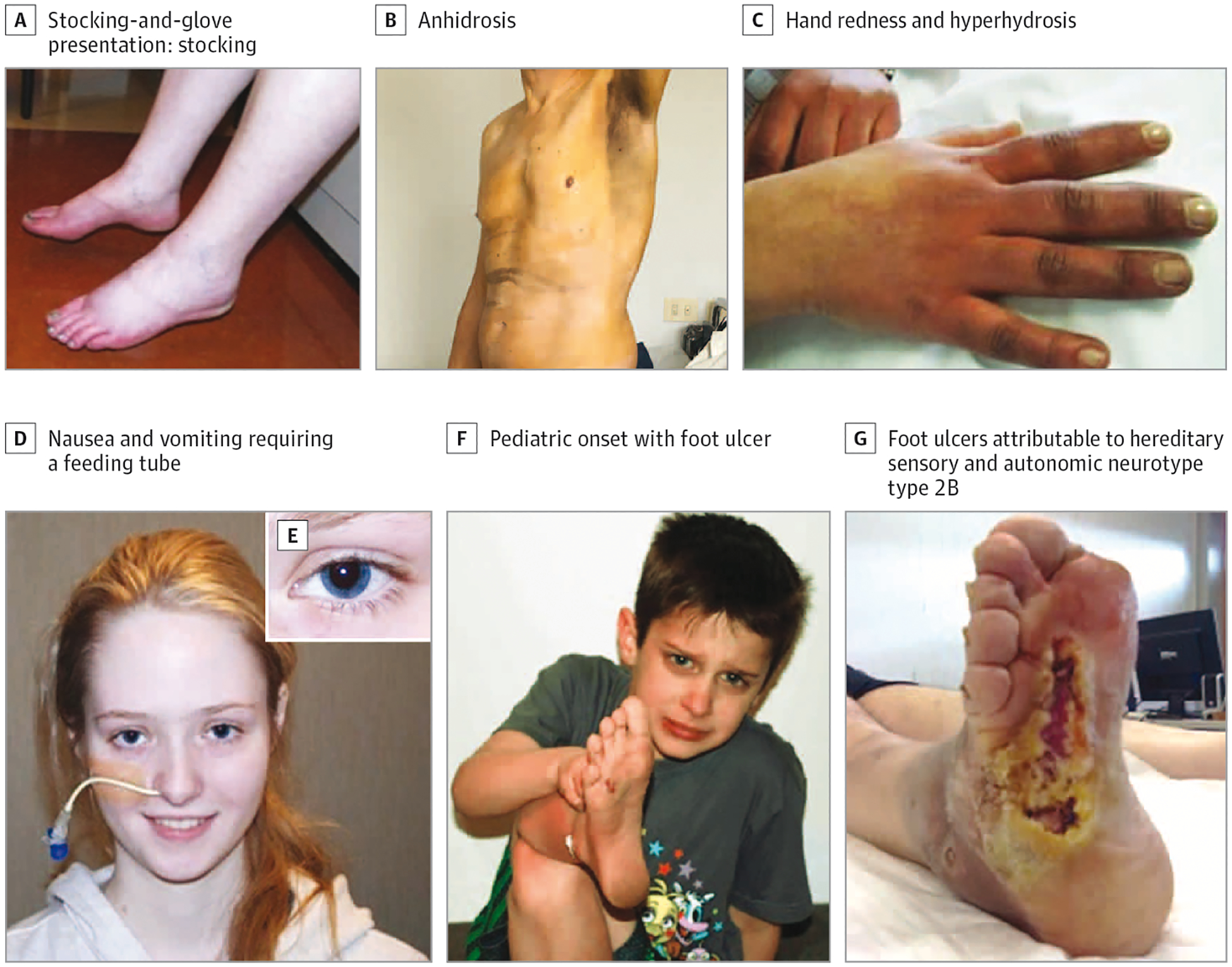
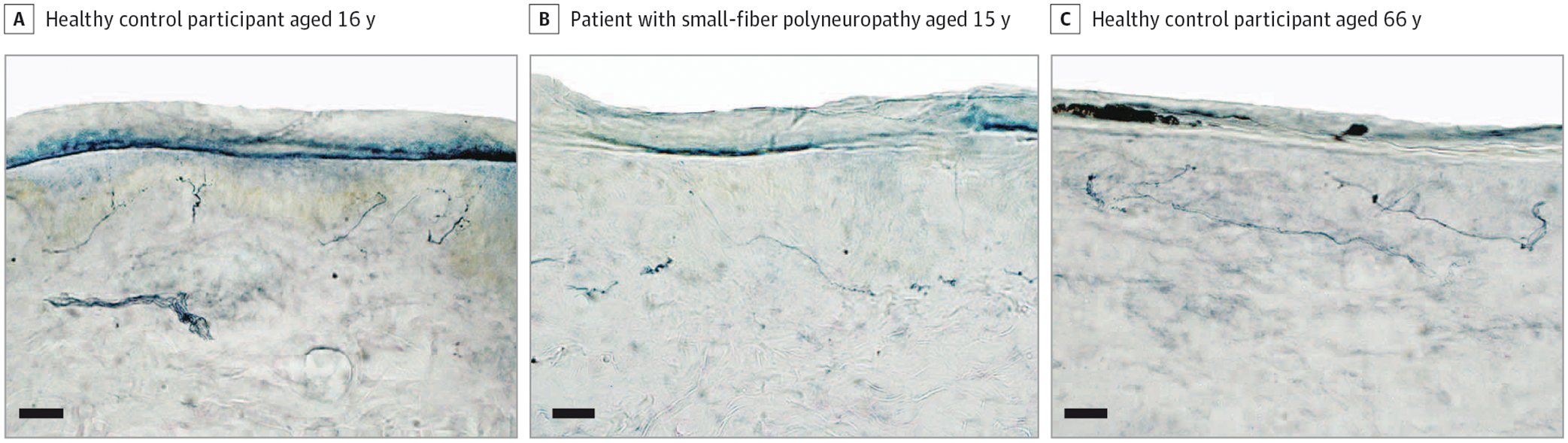
Observations: Biomarker confirmation is recommended, given the nonspecificity of symptoms. The standard test involves measuring epidermal neurite density within a 3-mm protein gene product 9.5 (PGP9.5)-immunolabeled lower-leg skin biopsy. Biopsies and autonomic function testing confirm that small-fiber neuropathy not uncommonly affects otherwise healthy children and young adults, in whom it is often associated with inflammation or dysimmunity. A recent meta-analysis concluded that small-fiber neuropathy underlies 49% of illnesses labeled as fibromyalgia. Initially, patients with idiopathic small-fiber disorders should be screened by medical history and blood tests for potentially treatable causes, which are identifiable in one-third to one-half of patients. Then, secondary genetic testing is particularly important for familial and childhood cases. Treatable genetic causes include Fabry disease, transthyretin and primary systemic amyloidosis, hereditary sensory autonomic neuropathy-1, and ion-channel mutations. Immunohistopathologic evidence suggests that small-fiber dysfunction and denervation, especially of blood vessels, contributes to diverse symptoms, including postexertional malaise, postural orthostatic tachycardia, and functional gastrointestinal distress. Preliminary evidence implicates acute or chronic autoreactivity in some cases, particularly in female patients and otherwise healthy children and young adults. Different temporal patterns akin to Guillain-Barré syndrome and chronic inflammatory demyelinating polyneuropathy have been described; here, corticosteroids and immunoglobulins, which are often efficacious for inflammatory neuropathic conditions, are increasingly considered.

Conclusions and relevance: Because small fibers normally grow throughout life, improving contributory conditions may permit regrowth, slow progression, and prevent permanent damage. The prognosis is often hopeful for improving quality of life and sometimes for abatement or resolution, particularly in the young and otherwise healthy individuals. Examples include diabetic, infectious, toxic, genetic, and inflammatory causes. The current standard of care requires prompt diagnosis and treatment, particularly in children and young adults, to restore life trajectory. Consensus diagnostic and tracking metrics should be established to facilitate treatment trials.


Conflict of interest statement

Conflict of Interest Disclosures: None reported.

Figures

Figure 1.
Figure 1.
Multiple Functions of Normal Cutaneous Small Fibers Reduced in a Patient With Early-Onset Small-Fiber Polyneuropathy Multilabel fluorescent immunohistochemistry with confocal microscopy. Axons are green (PGP 9.5), basement membrane and blood vessels are red (col IV), and epidermis and endothelia are blue (Ulex europaeus agglutinin). A, C, E, and G are from the skin biopsy of a normal control (a boy aged 10 years); B, D, F, and G are from a boy with Fabry disease (aged 13 years). Images are from the standard lower-leg site, except C and D, which are from a glabrous fingertip. A and B, Epidermal neurites; B shows morphological signs of degeneration (axon thinning and beading) and regeneration (clusters within tracts of epidermal denervation) and a disordered subepidermal neural plexus. C and D, Normal abundant innervation of arteriovenous anastomoses by vasomotor fibers to maintain tonic closure, which is severely reduced in D. E and F, Sudomotor innervation of sweat glands; F shows severe axonal denervation and derangement with resultant atrophy of the sweat gland. G and H, Pilomotor innervation of arrector pili muscles; the density is moderately reduced in H.
Figure 2.
Figure 2.
Neurological Examination Findings A, Classic stocking-and-glove erythromelalgia presentation in the distal extremities, with burning pain and neuropathic microvasculopathy causing skin redness and underlying edema. Severely affected patients often cool the affected skin to reduce C-fiber firing and pain. B, Non–length-dependent anhidrosis in a patient with small-fiber neuropathy from transthyretin amyloidosis. Thermoregulatory sweat testing results reveal residual sweating (dark staining) only in the axilla. C, Episodic hand redness, pain, and hyperhidrosis in woman with early-onset idiopathic small-fiber polyneuropathy. D and E, An adolescent with early-onset small-fiber polyneuropathy causing profound nausea, vomiting, and cachexia improved with a feeding tube (D); the same individual displays Adie pupils (E). F, A boy with biopsy-confirmed SFN attributable to SCN9A mutation cries from chronic neuropathic pain and demonstrates his lower-leg skin thickening and early painless foot ulcer from scratching neuropathic itch. G, A man with hereditary sensory and autonomic neuropathy type 2B from a RAB7 mutation has severe painless foot ulcers.
Figure 3.
Figure 3.
Protein Gene Product 9.5–Immunolabeled Lower-Leg Skin Biopsies: Importance of Accurate Norms for Diagnostic Interpretation This bright-field photomicrograph illustrates standard clinical processing and morphometric evaluations used most often for clinical diagnostic confirmation of small-fiber neuropathy. Skin biopsies are from 10 cm above the lateral malleolus in adults and proportionally less in children. They are cryosectioned vertically into 50-μm sections and hand-immunolabeled using monoclonal antibody targeting protein gene product–9.5, a ubiquitin hydrolase present in all axons. Skilled morphometrists count the number of neurites that penetrate the dermal-epidermal junction and express epidermal neurite density per mm2 of skin-surface area. Each patient’s epidermal neurite density is statistically compared with the anticipated normal distribution calculated using within-laboratory measurements made from biopsies of screened healthy control participants. Patient with epidermal neurite densities at less than the fifth percentile of expected amounts have pathologically confirmed clinical diagnoses, and predegenerative swellings, fragmentation, or inflammatory infiltrates (not shown) can be supportive; × 40 magnification; scale bar = 100 μm. A, Screened normal, healthy, white male control patient, aged 16 years. His epidermal neurite density (356 per mm2 of skin-surface area) was at the 58.0 percentile of the anticipated normal distribution of his age, sex, and race and provides no pathological evidence of small-fiber polyneuropathy. B, A 15.1-year-old white male patient with gastrointestinal symptoms, headache, fatigue, labile blood pressure, and postural orthostatic tachycardia syndrome (POTS) starting at age 3 years. His epidermal neurite density of 164 per mm2 of skin-surface area is at the 1.3rd percentile of the anticipated norm by age, sex, and race, confirming the clinician’s diagnosis of small-fiber polyneuropathy. Autonomic function testing results were also abnormal, antinuclear antibody test results were positive at 1:160, and a paraneoplastic panel identified autoantibodies against a voltage-gated potassium channel. C, This screened normal, healthy control participant, a 66.3-year-old white woman had 158 epidermal neurites per mm2 of skin-surface area and was at the 37.6th percentile of anticipated normal distribution for her age, sex, and race, well within the normal range, although lower than the pathological epidermal neurite density in B.

References

    1. Gregg EW, Sorlie P, Paulose-Ram R, et al. ; 1999–2000 National Health and Nutrition Examination Survey. Prevalence of lower-extremity disease in the US adult population ≥40 years of age with and without diabetes: 1999–2000 National Health and Nutrition Examination Survey. Diabetes Care. 2004;27(7):1591–1597. doi: 10.2337/diacare.27.7.1591 - DOI - PubMed
    1. Peters MJ, Bakkers M, Merkies IS, Hoeijmakers JG, van Raak EP, Faber CG. Incidence and prevalence of small-fiber neuropathy: a survey in the Netherlands. Neurology. 2013;81(15):1356–1360. doi: 10.1212/WNL.0b013e3182a8236e - DOI - PubMed
    1. Grayston R, Czanner G, Elhadd K, et al. A systematic review and meta-analysis of the prevalence of small fiber pathology in fibromyalgia: implications for a new paradigm in fibromyalgia etiopathogenesis. Semin Arthritis Rheum. 2019;48 (5):933–940. doi: 10.1016/j.semarthrit.2018.08.003 - DOI - PubMed
    1. Branco JC, Bannwarth B, Failde I, et al. Prevalence of fibromyalgia: a survey in five European countries. Semin Arthritis Rheum. 2010; 39(6):448–453. doi: 10.1016/j.semarthrit.2008.12.003 - DOI - PubMed
    1. Fridman V, Suriyanarayanan S, Novak P, et al. Randomized trial of l-serine in patients with hereditary sensory and autonomic neuropathy type 1. Neurology. 2019;92(4):e359–e370. doi: 10.1212/WNL.0000000000006811 - DOI - PMC - PubMed