

Glucocorticoid stimulation increases cardiac contractility by SGK1-dependent SOCE-activation in rat cardiac myocytes
- PMID: 31498847
- PMCID: PMC6733454
- DOI: 10.1371/journal.pone.0222341
Glucocorticoid stimulation increases cardiac contractility by SGK1-dependent SOCE-activation in rat cardiac myocytes
Abstract
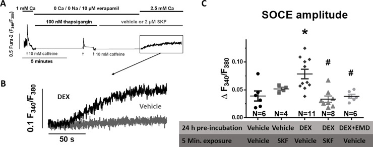
Aims: Glucocorticoid (GC) stimulation has been shown to increase cardiac contractility by elevated intracellular [Ca] but the sources for Ca entry are unclear. This study aims to determine the role of store-operated Ca entry (SOCE) for GC-mediated inotropy.
Methods and results: Dexamethasone (Dex) pretreatment significantly increased cardiac contractile force ex vivo in Langendorff-perfused Sprague-Dawley rat hearts (2 mg/kg BW i.p. Dex 24 h prior to experiment). Moreover, Ca transient amplitude as well as fractional shortening were significantly enhanced in Fura-2-loaded isolated rat ventricular myocytes exposed to Dex (1 mg/mL Dex, 24 h). Interestingly, these Dex-dependent effects could be abolished in the presence of SOCE-inhibitors SKF-96356 (SKF, 2 μM) and BTP2 (5 μM). Ca transient kinetics (time to peak, decay time) were not affected by SOCE stimulation. Direct SOCE measurements revealed a negligible magnitude in untreated myocytes but a dramatic increase in SOCE upon Dex-pretreatment. Importantly, the Dex-dependent stimulation of SOCE could be blocked by inhibition of serum and glucocorticoid-regulated kinase 1 (SGK1) using EMD638683 (EMD, 50 μM). Dex preincubation also resulted in increased mRNA expression of proteins involved in SOCE (stromal interaction molecule 2, STIM2, and transient receptor potential cation channels 3/6, TRPC 3/6), which were also prevented in the presence of EMD.
Conclusion: Short-term GC-stimulation with Dex improves cardiac contractility by a SOCE-dependent mechanism, which appears to involve increased SGK1-dependent expression of the SOCE-related proteins. Since Ca transient kinetics were unaffected, SOCE appears to influence Ca cycling more by an integrated response across multiple cardiac cycles but not on a beat-to-beat basis.
Conflict of interest statement
The authors have declared that no competing interests exist.
Figures


Similar articles
-
Transient Receptor Potential Canonical (TRPC)/Orai1-dependent Store-operated Ca2+ Channels: NEW TARGETS OF ALDOSTERONE IN CARDIOMYOCYTES.J Biol Chem. 2016 Jun 17;291(25):13394-409. doi: 10.1074/jbc.M115.693911. Epub 2016 Apr 22. J Biol Chem. 2016. PMID: 27129253 Free PMC article.
-
CaMKIIδ meditates phenylephrine induced cardiomyocyte hypertrophy through store-operated Ca2+ entry.Cardiovasc Pathol. 2017 Mar-Apr;27:9-17. doi: 10.1016/j.carpath.2016.11.004. Epub 2016 Dec 2. Cardiovasc Pathol. 2017. PMID: 27940402
-
Conformation of ryanodine receptor-2 gates store-operated calcium entry in rat pulmonary arterial myocytes.Cardiovasc Res. 2016 Jul 1;111(1):94-104. doi: 10.1093/cvr/cvw067. Epub 2016 Mar 24. Cardiovasc Res. 2016. PMID: 27013634 Free PMC article.
-
Tissue Specificity: Store-Operated Ca2+ Entry in Cardiac Myocytes.Adv Exp Med Biol. 2017;993:363-387. doi: 10.1007/978-3-319-57732-6_19. Adv Exp Med Biol. 2017. PMID: 28900924 Review.
-
Tuning store-operated calcium entry to modulate Ca2+-dependent physiological processes.Biochim Biophys Acta Mol Cell Res. 2019 Jul;1866(7):1037-1045. doi: 10.1016/j.bbamcr.2018.11.018. Epub 2018 Dec 3. Biochim Biophys Acta Mol Cell Res. 2019. PMID: 30521873 Review.
Cited by
-
A non-coding GWAS variant impacts anthracycline-induced cardiotoxic phenotypes in human iPSC-derived cardiomyocytes.Nat Commun. 2022 Nov 22;13(1):7171. doi: 10.1038/s41467-022-34917-y. Nat Commun. 2022. PMID: 36418322 Free PMC article.
-
Major surgery leads to a proinflammatory phenotype: Differential gene expression following a laparotomy.Ann Med Surg (Lond). 2021 Oct 21;71:102970. doi: 10.1016/j.amsu.2021.102970. eCollection 2021 Nov. Ann Med Surg (Lond). 2021. PMID: 34745602 Free PMC article.
-
Specific Upregulation of TRPC1 and TRPC5 Channels by Mineralocorticoid Pathway in Adult Rat Ventricular Cardiomyocytes.Cells. 2019 Dec 23;9(1):47. doi: 10.3390/cells9010047. Cells. 2019. PMID: 31878108 Free PMC article.
-
Mechanisms and Clinical Applications of Glucocorticoid Steroids in Muscular Dystrophy.J Neuromuscul Dis. 2021;8(1):39-52. doi: 10.3233/JND-200556. J Neuromuscul Dis. 2021. PMID: 33104035 Free PMC article. Review.
-
Biomimetic cardiac tissue culture model (CTCM) to emulate cardiac physiology and pathophysiology ex vivo.Commun Biol. 2022 Sep 9;5(1):934. doi: 10.1038/s42003-022-03919-3. Commun Biol. 2022. PMID: 36085302 Free PMC article.
References
Publication types
MeSH terms
Substances
LinkOut - more resources
Full Text Sources
Medical
Miscellaneous
