. 2019 Nov 1;384(1):111616.
doi: 10.1016/j.yexcr.2019.111616. Epub 2019 Sep 6.

Characterization of stitch adhesions: Fibronectin-containing cell-cell contacts formed by fibroblasts

Affiliations

Characterization of stitch adhesions: Fibronectin-containing cell-cell contacts formed by fibroblasts

Roumen Pankov et al. Exp Cell Res. .

Abstract

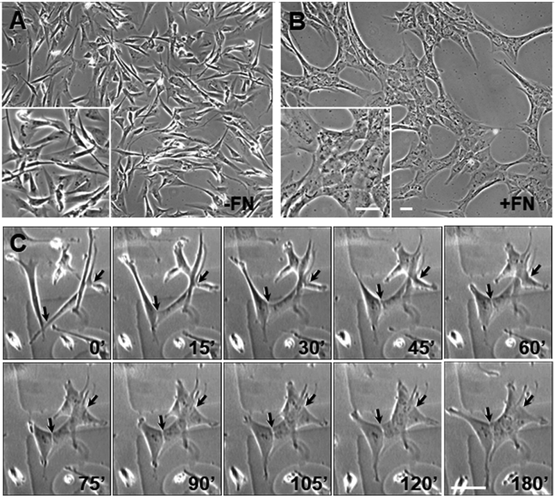
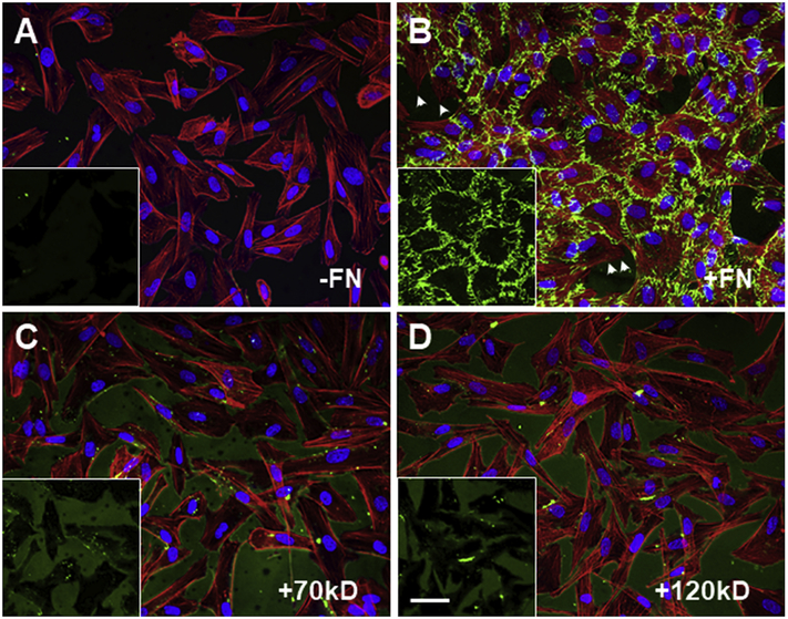
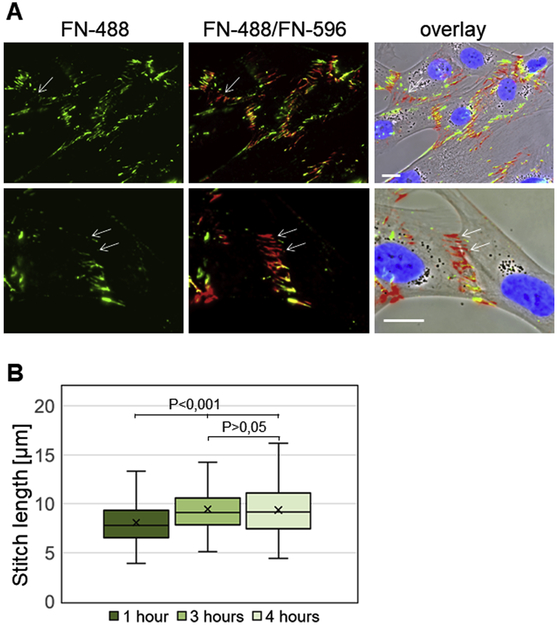
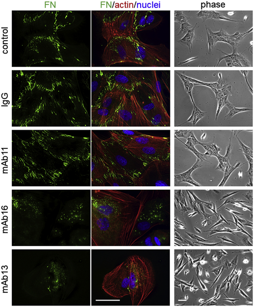
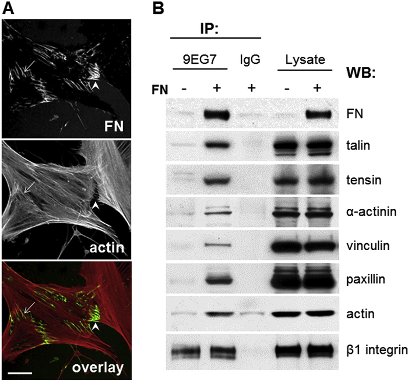
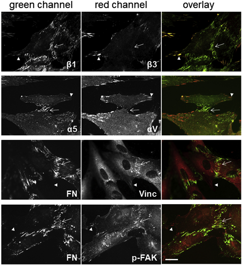
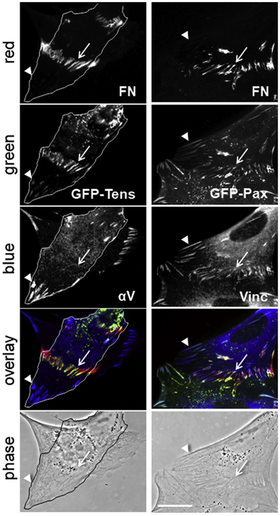
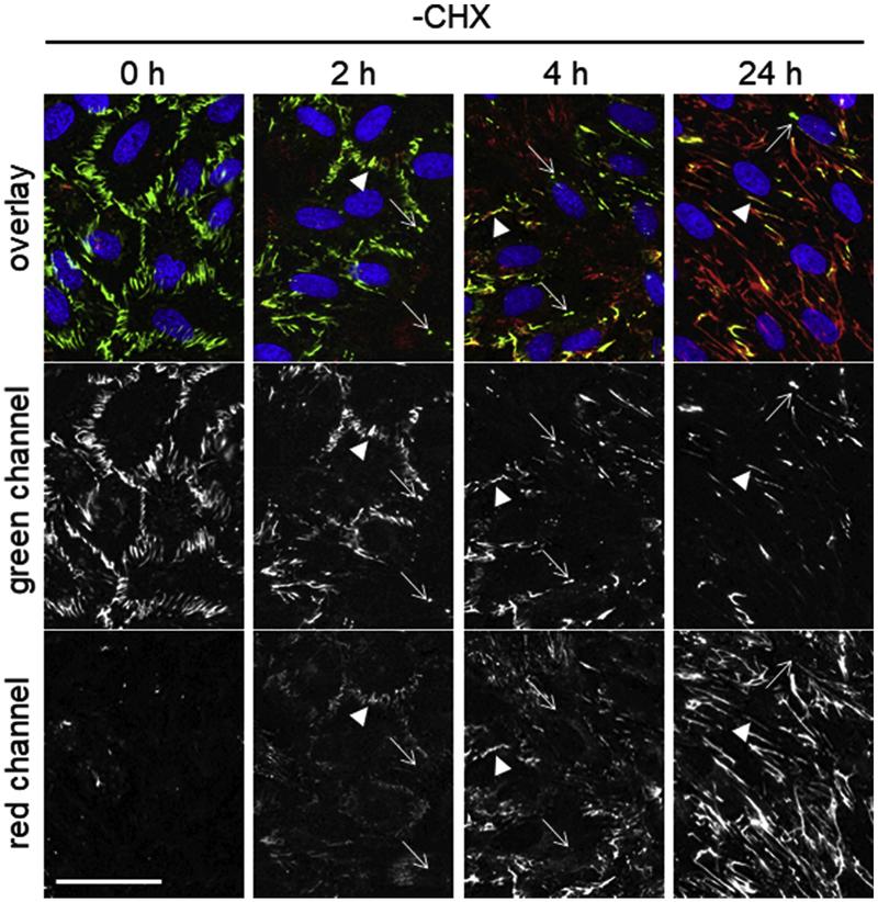
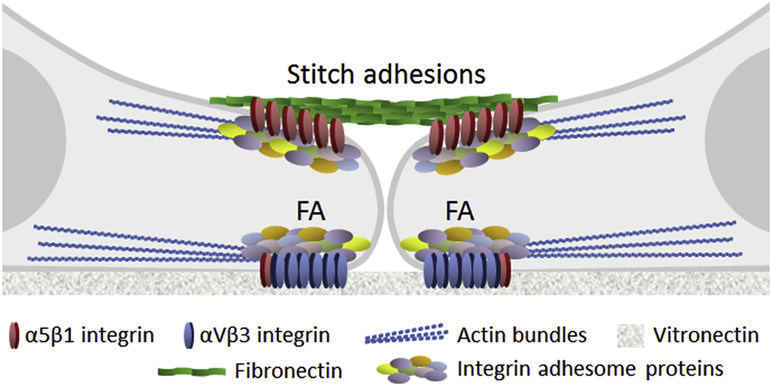
Fibronectin is a multifunctional, extracellular matrix glycoprotein that exists either as an insoluble multimeric fibrillar component of the extracellular matrix or as a soluble monomer. Cells attach to fibronectin through transmembrane integrin receptors and form a variety of cell-matrix contacts. Here we show that primary fibroblasts can use fibronectin to organize a specific cell-cell contact - "stitch adhesions." This contact is formed by short parallel fibronectin fibrils connecting adjacent cells above the level of the focal adhesions that attach the cells to the substrate. Stitch adhesions contain integrin α5β1 but not αVβ3, align with actin filament bundles, and contain talin, tensin, α-actinin, vinculin, paxillin and a phosphorylated form of focal adhesion kinase. This combination of components differs from the described constituents of the known cell adhesions. Stitch adhesions are organized when protein synthesis and secretion are inhibited by cycloheximide and exogenous fibronectin is provided to the cells. The adhesion stitches described here provide an attractive model system for studying fibronectin fibrillogenesis and the mechanisms governing the formation of cellular adhesions.

Keywords: Adhesion; Cell-cell contacts; Cycloheximide; Fibronectin; Human fibroblasts.


Figures

Figure 1.
Figure 1.
Full-length fibronectin induction of formation of cell-to-cell contacts in cycloheximide-treated fibroblasts. Phase-contrast images of CHX-treated cells revealed elongated morphologies in the absence of FN (A), but after incubation with 25 μg/ml human plasma FN for 4 hours (B), they developed extended cell-cell contacts, combined with an epithelial-like morphology. Time-lapse images acquired at 15 min intervals demonstrated quick initial establishment of cell-to-cell contacts that gradually increased (C). Arrows indicate developing cell-cell contacts. Bar = 50μm.
Figure 2.
Figure 2.
Immunofluorescence of CHX-treated fibroblasts incubated with full length FN or fragments of FN and stained for fibronectin (green) and actin (red). (A) The control CHX-treated cells did not stain with anti-fibronectin antibodies, indicating that synthesis and secretion of FN were completely blocked. (B) Prominent fibronectin stitches were observed between adjacent cells after incubation with full-length plasma FN. No organized FN was detected in samples incubated for the same period of time with 70kD (C) or 120kD (D) fibronectin fragments. Nuclei (blue) were stained with Hoechst. Insets present only fibronectin staining. Arrowheads in B indicate free cellular edges devoid of FN. Bar = 50μm.
Figure 3.
Figure 3.
Dynamics of FN stitch development. (A) Pulse-chase analysis of the formation of stitch adhesions using labeled fibronectin preparations. CHX-treated fibroblasts were incubated with fibronectin labeled with green Alexa 488 (FN-488) for 1 hour, followed by another hour of incubation with fibronectin labeled with red Alexa 594 (FN-488/FN-594). Arrows indicate stitches formed during the second hour of incubation. Merged fluorescence and phase contrast images show localization of labeled stitches at places of cell-cell contacts. Nuclei were visualized with Hoechst (blue). Bar = 20μm. (B) Determination of stitch length. CHX-treated fibroblast were incubated with fibronectin for different periods, fixed and stained with anti-fibronectin antibodies. The lengths of FN stitches were measured on immunofluorescence images and presented as box plots.
Figure 4.
Figure 4.
Blocking integrin α5β1 prevents the formation of StA and cell-to-cell contacts. Cycloheximide-treated human foreskin fibroblasts were incubated for 30 minutes with 200 μg/ml IgG2a,κ isotype antibody control (IgG), nonfunction-blocking anti-α5 antibody (mAb11), inhibitory anti-α5 antibody (mAb16), and inhibitory anti-β1antibody (mAb13) or were left untreated (control). Stitch adhesions were induced in all samples by incubation with 25 μg/ml HiLyte Fluor™ 488-labeled bovine FN in presence of the same concentrations of antibodies for an additional 4 hours. Fibronectin was observed using the green fluorescence channel (FN), rhodamine-phalloidin was used for visualization of the actin cytoskeleton (red channel), and nuclei were stained with Hoechst (blue channel). Merged images with the corresponding colors are shown under “FN/actin/nuclei”. General morphology of the cells was determined using phase contrast microscopy (phase). Bar = 50μm.
Figure 5.
Figure 5.
Adhesion stitches aligned with actin filament bundles and associated with proteins typical of the integrin adhesome. Immunofluorescence images of (A) CHX-treated fibroblasts incubated with 25 μg/ml human plasma FN for 4 hours and then stained with anti-fibronectin antibodies (FN) and rhodamine-phalloidin (actin). Merged images (overlay) demonstrated alignment between actin bundles and fibronectin stitches (arrows). Fibronectin fibrils occasionally bridged the gap between neighboring cells (arrowheads). Bar = 20μm. Co-immunoprecipitation experiments of (B) CHX-treated cells in the absence (−FN) or presence (+FN) of fibronectin performed with anti-β1 integrin antibody 9EG7 (IP:9EG7) and rat IgG2a,κ isotype antibody control (IP:IgG), followed by Western blotting (WB) with antibodies against the indicated proteins revealed strong enrichment of adhesome proteins in β1 integrin complexes after incubation of cells with FN. Loading controls prior to immunoprecipitation (Lysate) are also shown.
Figure 6.
Figure 6.
Identification of molecular components of the stitch adhesions. Cycloheximide-treated human foreskin fibroblasts were incubated with fibronectin and stained with antibodies against the indicated integrins, adhesome proteins and FN. The primary antibodies were visualized with secondary antibodies conjugated to FITC (green channel) or Cy3 (red channel). Merged images are presented in the righthand column (overlay). Arrows indicate stitch adhesions, and arrowheads focal adhesions. Bar = 20μm.
Figure 7.
Figure 7.
GFP-tagged tensin and paxillin localize at stitch adhesions. Transiently transfected HFF with GFP-tensin (left column) and GFP-paxillin (right column) were stained with antibodies against fibronectin (FN), αV integrin (αV) and vinculin (Vinc). The primary antibodies were visualized with secondary antibodies conjugated to Cy3 (red) or AMCA (blue). GFP fluorescence was detected on the green channel (green). Merged images are presented under “overlay” and phase contrast images under “phase”. Arrows indicate stitch adhesions and arrowheads - focal adhesions. The cell positioned closer to the substrate in the images in the left column is outlined. Bar = 20μm.
Figure 8.
Figure 8.
Removal of cycloheximide restores normal fibronectin fibrillogenesis. (A) Fibroblasts treated (+CHX) or not treated (−CHX) with cycloheximide overnight in media containing 1% FN-depleted serum were incubated (+bFN) or not (−bFN) with 25 μg/ml HiLyte Fluor™ 488-labeled bovine FN for 4 hours. All samples were washed and left overnight in the above medium in the absence of both cycloheximide and labeled FN (O.N. in medium −CHX −bFN). To visualize the endogenously produced human fibronectin, samples were stained with anti-human FN antibody and secondary Cy3-conjugated antibody. The green channel was used to detect bovine FN (bFN), the red channel for human FN (hFN), and the blue channel for Hoechst-stained nuclei (Hoechst). Arrowheads indicate hybrid FN fibers containing both bovine and human fibronectin; arrows indicate FN fibers originating from StA. (B) Fibroblasts treated (+CHX) or not treated (−CHX) with cycloheximide as above were grown in media containing 1% FN-depleted serum for an additional 5 days. Fibronectin matrix was stained with anti-human FN antibody (red) and nuclei were stained with Hoechst (blue). Bar = 50μm.
Figure 9.
Figure 9.
Reorganization of stitch adhesions after restoration of normal fibronectin fibrillogenesis. Fibroblasts with organized StA (0 h) were washed and incubated in CHX-free DMEM containing 1% FN-depleted serum for the indicated times. Stitch adhesions formed by HiLyte Fluor™ 488-labeled bovine FN were detected in the green channel and the secreted human FN was observed in the red channel after staining with anti-human FN antibody and secondary Cy3-conjugated antibody. Nuclei were stained with Hoechst. Merged fluorescent images are presented under “overlay”. Arrowheads indicate hybrid FN fibers containing bovine and human fibronectin, and arrows indicate – FN fibers originating from StA. Bar = 50μm.
Figure 10.
Figure 10.
Schematic representation of the stitch adhesion. Unlike focal adhesions (FA), which are located at the ventral cell surface and attach cells to the substrate mainly through the vitronectin receptor αVβ3, stitch adhesions are positioned dorsally and connect adjacent cells by short fibronectin fibrils bound exclusively to α5β1 integrins. Both types of adhesions connect to the actin cytoskeleton through protein plaques composed of similar members of the integrin adhesome.

Similar articles

Cited by

References

    1. Hynes RO, Fibronectins. New York: Springer-Verlag, 1990. http://www.springer.com/la/book/9781461279402
    1. Pankov R, Yamada KM, Fibronectin at a glance, J. Cell Sci. 115 (2002) 3861–3863. http://jcs.biologists.org/content/115/20/3861.long - PubMed
    1. Singh P, Carraher C, Schwarzbauer JE, Assembly of fibronectin extracellular matrix, Annu. Rev. Cell Dev. Biol. 26 (2010) 397–419. https://www.annualreviews.org/doi/abs/10.1146/annurev-cellbio-100109-104020 - DOI - PMC - PubMed
    1. George EL, Georges-Labouesse EN, Patel-King RS, Rayburn H, Hynes RO, Defects in mesoderm, neural tube and vascular development in mouse embryos lacking fibronectin, Development, 119 (1993) 1079–1091. http://dev.biologists.org/content/119/4/1079.short - PubMed
    1. Plow EF, Haas TA, Zhang L, Loftus J, Smith JW, Ligand binding to integrins, J. Biol. Chem. 275 (2000) 21785–21788. http://www.jbc.org/content/275/29/21785.short - PubMed

Publication types