. 2019 Sep 6;8(9):1046.
doi: 10.3390/cells8091046.

Viscosity of Plasma as a Key Factor in Assessment of Extracellular Vesicles by Light Scattering

Affiliations

Viscosity of Plasma as a Key Factor in Assessment of Extracellular Vesicles by Light Scattering

Darja Božič et al. Cells. .

Abstract

Extracellular vesicles (EVs) isolated from biological samples are a promising material for use in medicine and technology. However, the assessment methods that would yield repeatable concentrations, sizes and compositions of the harvested material are missing. A plausible model for the description of EV isolates has not been developed. Furthermore, the identity and genesis of EVs are still obscure and the relevant parameters have not yet been identified. The purpose of this work is to better understand the mechanisms taking place during harvesting of EVs, in particular the role of viscosity of EV suspension. The EVs were harvested from blood plasma by repeated centrifugation and washing of samples. Their size and shape were assessed by using a combination of static and dynamic light scattering. The average shape parameter of the assessed particles was found to be ρ ~ 1 (0.94-1.1 in exosome standards and 0.7-1.2 in blood plasma and EV isolates), pertaining to spherical shells (spherical vesicles). This study has estimated the value of the viscosity coefficient of the medium in blood plasma to be 1.2 mPa/s. It can be concluded that light scattering could be a plausible method for the assessment of EVs upon considering that EVs are a dynamic material with a transient identity.

Keywords: blood plasma; dynamic light scattering; exosomes; extracellular vesicles; shape characterization; static light scattering; viscosity of blood plasma.


Conflict of interest statement

The authors declare no conflicts of interest.

Figures

Figure 1
Figure 1
Atomic force microscopy (AFM) micrographs of (A) an unfiltered, (B) filtered exosome standard, (C) an extracellular vesicle (EV) isolate from a blood plasma sample, and (D) magnification of the area indicated in C.
Figure 2
Figure 2
(A) The measured correlation functions G2(t) and (B) the calculated size distributions of particles in an unfiltered exosome standard (solid black line), a fresh plasma sample (dashed red line) and the corresponding EV isolate from the same sample (dotted black line). In calculation of Rh (Equation (2)) for the exosome standard, the viscosity coefficient of water (η0 = 0.9 mPa/s) was used, while for the plasma sample both, water (dashed red line) and the estimated viscosity of the plasma medium (η = 1.2 mPa/s; double red line; for details see text below) were taken into account. The arrows depict the shift of peak positions in the plasma sample after the viscosity correction.
Figure 3
Figure 3
The intensity of scattered light measured by static light scattering (SLS) and represented as the Kratky plot (i.e., (qRg)2P(q) versus qRg) for the population 2 (large particles; see text) in the exosome standard ES (open circles: unfiltered; full circles: filtered), a plasma sample (full triangles) and the corresponding EV isolate from the same sample (open triangles). The lines show the calculated (qRg)2P(q) versus qRg dependencies for some selected shapes (for details see Supplementary Material).
Figure 4
Figure 4
The AF4/UV-MALS fractograms of unfiltered (black) and filtered (red) exosome standards (ES) together with radius of gyration Rg as a function of time for smaller (open circles) and larger (full circles) vesicle populations. The solid lines represent the LS detector responses at 90° angle, while the dashed curves represent the UV detector responses.
Figure 5
Figure 5
Kratky plots (qRg)2P(q) vs. qRg) were constructed for large particles eluted at a peak apex (32 min, full circles) and at ~35 min (open circles) in the AF4/UV-MALS fractogram of the filtered exosome standard (ES).
Figure 6
Figure 6
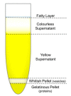
The fractions of the blood plasma sample after 8 h of ultracentrifugation at 100,000× g.

References

    1. Simpson R.J., Mathivanan S. Extracellular Microvesicles: The Need for Internationally Recognised Nomenclature and Stringent Purification Criteria. J. Proteomics Bioinform. 2012 doi: 10.4172/jpb.10000e10. - DOI
    1. Linares R., Tan S., Gounou C., Arraud N., Brisson A.R. High-speed centrifugation induces aggregation of extracellular vesicles. J. Extracell. Vesicles. 2015 doi: 10.3402/jev.v4.29509. - DOI - PMC - PubMed
    1. Johnsen K.B., Gudbergsson J.M., Andresen T.L., Simonsen J.B. What is the blood concentration of extracellular vesicles? Implications for the use of extracellular vesicles as blood-borne biomarkers of cancer. Biochim. Biophys. Acta. 2019;1871:109–116. doi: 10.1016/j.bbcan.2018.11.006. - DOI - PubMed
    1. Černe K., Kobal B. Chapter Eight-Implications of Microvesicle and Cell Surface Protein Shedding for Biomarker Studies, Cancerogenesis, and Therapeutic Target Discovery in Ovarian Cancer. In: Aleš I., editor. Advances in Planar Lipid Bilayers and Liposomes. Volume 16. Academic Press; Cambridge, MA, USA: 2012. pp. 239–274.
    1. Yáñez-Mó M., Siljander P.R.M., Andreu Z., Zavec A.B., Borràs F.E., Buzas E.I., Buzas K., Casal E., Cappello F., Carvalho J., et al. Biological properties of extracellular vesicles and their physiological functions. J. Extracell. Vesicles. 2015 doi: 10.3402/jev.v4.27066. - DOI - PMC - PubMed

Publication types

LinkOut - more resources