. 2019 Sep 6;20(18):4394.
doi: 10.3390/ijms20184394.

Crystal Structures of Pyrophosphatase from Acinetobacter baumannii: Snapshots of Pyrophosphate Binding and Identification of a Phosphorylated Enzyme Intermediate

Affiliations

Crystal Structures of Pyrophosphatase from Acinetobacter baumannii: Snapshots of Pyrophosphate Binding and Identification of a Phosphorylated Enzyme Intermediate

Yunlong Si et al. Int J Mol Sci. .

Abstract

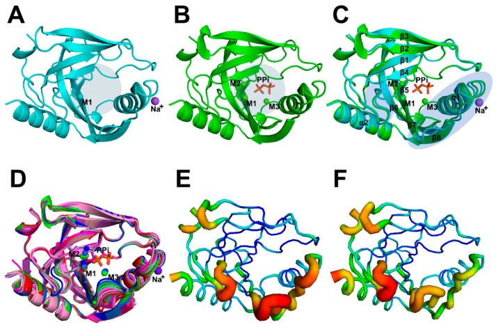
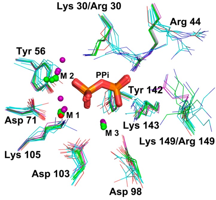
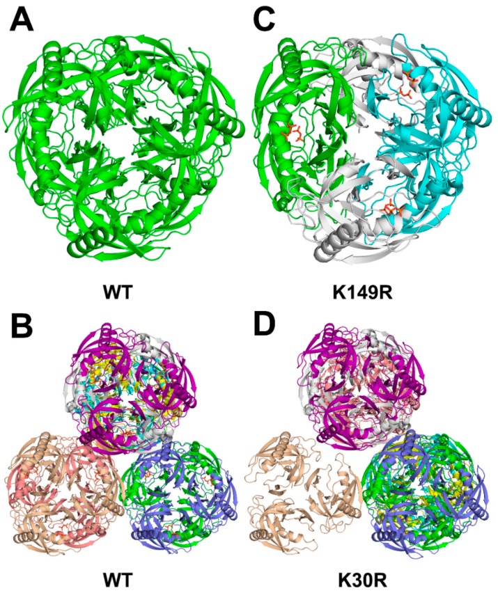
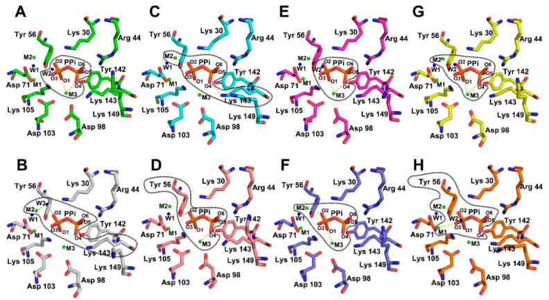
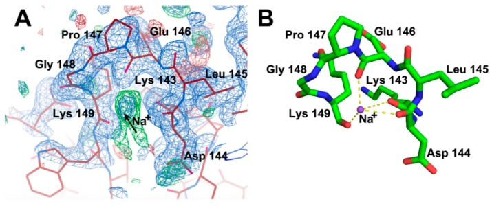
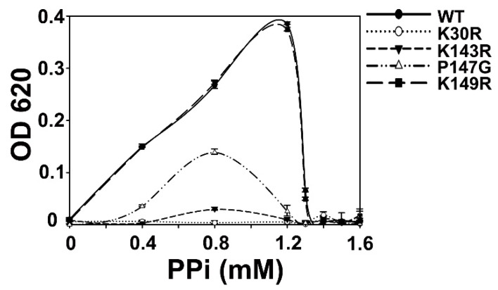
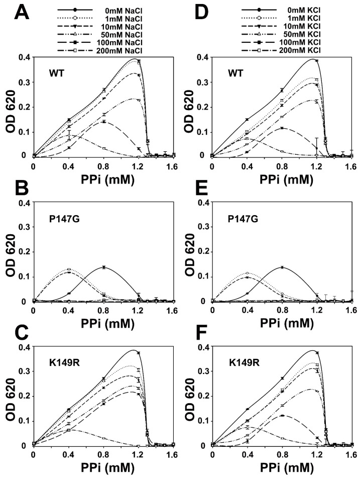
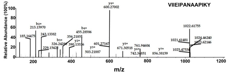
All living things have pyrophosphatases that hydrolyze pyrophosphate and release energy. This energetically favorable reaction drives many energetically unfavorable reactions. An accepted catalytic model of pyrophosphatase shows that a water molecule activated by two divalent cations (M1 and M2) within the catalytic center can attack pyrophosphate in an SN2 mechanism and thus hydrolyze the molecule. However, our co-crystal structure of Acinetobacter baumannii pyrophosphatase with pyrophosphate shows that a water molecule from the solvent may, in fact, be the actual catalytic water. In the co-crystal structure of the wild-type pyrophosphatase with pyrophosphate, the electron density of the catalytic centers of each monomer are different from one another. This indicates that pyrophosphates in the catalytic center are dynamic. Our mass spectroscopy results have identified a highly conserved lysine residue (Lys30) in the catalytic center that is phosphorylated, indicating that the enzyme could form a phosphoryl enzyme intermediate during hydrolysis. Mutation of Lys30 to Arg abolished the activity of the enzyme. In the structure of the apo wild type enzyme, we observed that a Na+ ion is coordinated by residues within a loop proximal to the catalytic center. Therefore, we mutated three key residues within the loop (K143R, P147G, and K149R) and determined Na+ and K+-induced inhibition on their activities. Compared to the wild type enzyme, P147G is most sensitive to these cations, whereas K143R was inactive and K149R showed no change in activity. These data indicate that monovalent cations could play a role in down-regulating pyrophosphatase activity in vivo. Overall, our results reveal new aspects of pyrophosphatase catalysis and could assist in the design of specific inhibitors of Acinetobacter baumannii growth.

Keywords: Acinetobacter baumannii; catalytic mechanism; crystal structure; inorganic pyrophosphatase; lysine phosphorylation; mass spectrometry.


Conflict of interest statement

The authors declare no conflict of interest.

Figures

Figure 1
Figure 1
Crystal structures and B-factors analysis of WT AbPPase. (A) Structure 1 of AbPPase without PPi. One Mg2+ (M1) bound to the catalytic center, and one Na+ is bound to a loop close to the catalytic center. Round light gray shades represent the catalytic center position. (B) Structure 2 of AbPPase with three Mg2+ ions (M1, M2, and M3) and one PPi bound at the catalytic center. Round light gray shades represent the catalytic center position. (C) Alignment of the Structures 1 and 2 of AbPPase. These structures are similar to each other. However, two regions close to the catalytic center could not be perfectly merged, i.e., region 1 located between β4 and β5 and region 2 located between α1 and β8. Elliptical light purple shades represent the positions of both loops. (D) Alignment all of the monomers from Structures 1, 2, 3, and 4. The monomer from Structure 1 of AbPPase with Mg2+ is marked in red, eight monomers from Structure 2 of the AbPPase with Mg2+ are marked in green. The eight monomers from Structure 3 (K30R) of the AbPPase are marked in magenta. The three monomers from Structure 4 (K149R) of AbPPase with Mg2+ are marked in blue. Two regions close to the catalytic center also could not be perfectly merged among different monomers. (E) B-factor analysis of Structure 1 of AbPPase. Two loops, including region 1 located between β4 and β5 and region 2 located between α1 and β8 that around the catalytic center are flexible. (F) B-factor analysis of one monomer of Structure 2.
Figure 2
Figure 2
Alignment of all catalytic centers of AbPPase monomers. Structure 1 of AbPPase without PPi was marked in red, Structure 2 of AbPPase with PPi was marked with green, Structure 3 (K30R) of AbPPase without PPi was marked with cyan, and Structure 4 (K149R) of AbPPase with PPi was marked in purple.
Figure 3
Figure 3
Top view of crystal packings of AbPPase. (A) Crystal structure of AbPPase without PPi, in which there is only one monomer in the asymmetric unit. Only one type of AbPPase hexamers was assembled. (B) Crystal structure of AbPPase with PPi, in which there are eight AbPPase monomers in the asymmetric unit. Eight different trimers could assemble four different types of AbPPase hexamers. (C) Crystal structure (K149R) of AbPPase with PPi in which there are three AbPPase monomers form a trimer in the asymmetric unit. One type of AbPPase hexamers was assembled. (D) Crystal structure (K30R) of AbPPase without PPi, in which there are eight AbPPase monomers in the asymmetric unit, and could assemble four different types of AbPPase hexamers and one type of trimer.
Figure 4
Figure 4
Variable PPi binding snapshots in eight AbPPase catalytic centers according to the distribution of electron densities. The dash lines delimit the regions where the electron densities of the atoms were fused with each other. Monomer (A,B), (C,D), (E,F), and (G,H) assemble into AbPPase hexamer 1, 2, 3, and 4. A water molecule (W2) from the solvent is close to the phosphate group and could be the real catalytic water (A). The left phosphate in PPi is named as Pi1 and the right one is named as Pi2.
Figure 5
Figure 5
Na+ was identified in crystal Structure 1 of AbPPase. The 2|Fo|–|Fc|, αc map contoured at 1δ is shown as blue density. The |Fo|–|Fc|, difference density map contoured at 3δ is shown as green density. (A) The electron density map of Na+ molecule in Structure 1 of AbPPase. Black arrow indicates where the Na+ molecule is located. (B) The coordination of Na+ molecule by a loop in Structure 1 of AbPPase. Lys143 and Lys149 make coordination with Na+ through their β-ketone groups. Pro147 forms the turn for this loop.
Figure 6
Figure 6
Enzyme assay of WT and four variants of AbPPase. The K149R variant had a similar activity as the WT AbPPase, K30R and K143R variants lost almost all their activity, and the P147G variant had a significantly reduced activity compared to the WT AbPPase. Error bars represent standard deviations for n = 3 independent experiments.
Figure 7
Figure 7
The enzyme assay inhibitory effects of Na+ and K+ on WT, P147G, and K149R of AbPPase. (A,D) Na+ and K+ could, in a concentration-dependent manner, inhibit WT of AbPPase hydrolysis of PPi, respectively. The higher the Na+ and K+ concentration, the lower the WT of AbPPase catalysis activity. (B,E) Na+ and K+ inhibit P147G of AbPPase hydrolysis of PPi, respectively. (C,F) Na+ and K+ could, in a concentration-dependent manner, inhibit K149R of AbPPase hydrolysis of PPi, respectively. K149R variant showed a similar behavior to the WT AbPPase to the inhibition of monovalent cations. Error bars represent standard deviation for n = 3 independent experiments.
Figure 8
Figure 8
Gel filtration analysis of the global fold of AbPPase. (A) Gel filtration profile of AbPPase in the presence of 1.6 mM PPi and 1.6 mM MgCl2. The elution peak of AbPPase falls at 13.36 mL. (B) The gel filtration profile of AbPPase in the presence of 200 mM NaCl,1.6 mM PPi and 1.6 mM MgCl2. The elution peak of AbPPase falls at 13.32 mL.
Figure 9
Figure 9
Mass spectrometry analysis of recombinant AbPPase after the in vitro PPi hydrolysis reaction to determine phosphorylation sites. A sufficient number of complementary Bn anions (N terminus-derived fragment ions) and Yn ions (C terminus-derived fragment ions) were detected to assign the phosphorylation sites to particular amino acids.

Similar articles

Cited by

References

    1. Nelson D., Cox M. Lehninger Principles of Biochemistry. 3rd ed. Worth Publishers; New York, NY, USA: 2000. p. 937.
    1. Reginald H., Charles M. Grisham Biochemistry. 4th ed. Cengage Learning, Brooks/Cole; Belmont, CA, USA: 2010.
    1. Carman G.M., Han G.S. Roles of phosphatidate phosphatase enzymes in lipid metabolism. Trends Biochem. Sci. 2006;31:694–699. doi: 10.1016/j.tibs.2006.10.003. - DOI - PMC - PubMed
    1. Van Wazer J.R., Griffith E.J., Mccullough J.F. Structure and Properties of the Condensed Phosphates. VII. Hydrolytic Degradation of Pyro- and Tripolyphosphate. J. Am. Chem. Soc. 1955;77:287–291. doi: 10.1021/ja01607a011. - DOI
    1. Sivula T., Salminen A., Parfenyev A.N., Pohjanjoki P., Goldman A., Cooperman B.S., Baykov A.A., Lahti R. Evolutionary aspects of inorganic pyrophosphatase. FEBS Lett. 1999;454:75–80. doi: 10.1016/S0014-5793(99)00779-6. - DOI - PubMed

LinkOut - more resources