Review
. 2019 Sep 8;11(9):1332.
doi: 10.3390/cancers11091332.

Bone Metastases in Neuroendocrine Neoplasms: From Pathogenesis to Clinical Management

Affiliations
Review

Bone Metastases in Neuroendocrine Neoplasms: From Pathogenesis to Clinical Management

Barbara Altieri et al. Cancers (Basel). .

Abstract

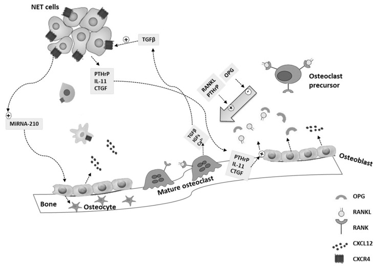
Bone represents a common site of metastases for several solid tumors. However, the ability of neuroendocrine neoplasms (NENs) to localize to bone has always been considered a rare and late event. Thanks to the improvement of therapeutic options, which results in longer survival, and of imaging techniques, particularly after the introduction of positron emission tomography (PET) with gallium peptides, the diagnosis of bone metastases (BMs) in NENs is increasing. The onset of BMs can be associated with severe skeletal complications that impair the patient's quality of life. Moreover, BMs negatively affect the prognosis of NEN patients, bringing out the lack of curative treatment options for advanced NENs. The current knowledge on BMs in gastro-entero-pancreatic (GEP) and bronchopulmonary (BP) NENs is still scant and is derived from a few retrospective studies and case reports. This review aims to perform a critical analysis of the evidence regarding the role of BMs in GEP- and BP-NENs, focusing on the molecular mechanisms underlining the development of BMs, as well as clinical presentation, diagnosis, and treatment of BMs, in an attempt to provide suggestions that can be used in clinical practice.

Keywords: bone metastases; bone microenvironment; denosumab; epithelial-to-mesenchymal transition; microRNA; neuroendocrine neoplasms; prognosis; skeletal-related events; treatment.


Conflict of interest statement

The authors declare no conflicts of interest.

Figures

Figure 1
Figure 1
Molecular mechanism of bone metastasis in neuroendocrine neoplasms: the “vicious cycle”. Neuroendocrine neoplasms (NENs) cells secrete pro-osteolytic factors, including the parathyroid hormone-related protein (PTHrP), interleukin-11 (IL-11), and the connective tissue growth factor (CTGF), which stimulate the activator receptor of the nuclear factor-kappa B ligand (RANKL) production by osteoblasts and/or the decrease of osteoprotegerin (OPG) within the bone stroma. Thus, RANKL induces osteoclast formation. Osteoclastic bone resorption causes the release and activation of growth factors, including the transforming growth factor-β (TGFβ), the insulin-like growth factors 1 (IGF1), and calcium ions (Ca2+). TGFβ can increase tumor production of the C-X-C motif receptor 4 (CXCR4) in NEN cells. The C-X-C motif chemokine-ligand-12 (CXCL12) is mainly produced by osteoblasts and can attract CXCR4-overexpressing NEN cells. CXCR4 and CTGF play a role in the migration of NEN cells to the bone. Micro-RNA (miRNA)-210 is upregulated in NEN cells and regulates the differentiation of osteoblasts into osteocytes.
Figure 2
Figure 2
Thoracic vertebrae (T4) bone metastasis (BM) was diagnosed two years after a liver metastasis in a 66-year-old woman with a history of ileal neuroendocrine neoplasm. Somatostatin receptor scintigraphy (SRS) was performed twice in the last four years of follow-up and did not detect BM. (a1,a2) T4 vertebra BM was first detected by 68Ga-DOTATOC positron emission tomography (PET)/ computed tomography (CT); (b1,b2) BM was confirmed by magnetic resonance imaging (MRI); (c) 99mTc-bone scintigraphy did not detect BM; (d) computed tomography was also negative for BM. Arrow indicates the BM.

References

    1. Faggiano A., Ferolla P., Grimaldi F., Campana D., Manzoni M., Davi M.V., Bianchi A., Valcavi R., Papini E., Giuffrida D., et al. Natural history of gastro-entero-pancreatic and thoracic neuroendocrine tumors. Data from a large prospective and retrospective Italian epidemiological study: The NET management study. J. Endocrinol. Invest. 2012;35:817–823. doi: 10.3275/8102. - DOI - PubMed
    1. Boyar Cetinkaya R., Aagnes B., Myklebust T.A., Thiis-Evensen E. Survival in neuroendocrine neoplasms; A report from a large Norwegian population-based study. Int. J. Cancer. 2018;142:1139–1147. doi: 10.1002/ijc.31137. - DOI - PubMed
    1. Dasari A., Shen C., Halperin D., Zhao B., Zhou S., Xu Y., Shih T., Yao J.C. Trends in the Incidence, Prevalence, and Survival Outcomes in Patients with Neuroendocrine Tumors in the United States. JAMA Oncol. 2017;3:1335–1342. doi: 10.1001/jamaoncol.2017.0589. - DOI - PMC - PubMed
    1. Hallet J., Law C.H., Cukier M., Saskin R., Liu N., Singh S. Exploring the rising incidence of neuroendocrine tumors: A population-based analysis of epidemiology, metastatic presentation, and outcomes. Cancer. 2015;121:589–597. doi: 10.1002/cncr.29099. - DOI - PubMed
    1. Barrea L., Altieri B., Muscogiuri G., Laudisio D., Annunziata G., Colao A., Faggiano A., Savastano S. Impact of Nutritional Status on Gastroenteropancreatic Neuroendocrine Tumors (GEP-NET) Aggressiveness. Nutrients. 2018;10:1854. doi: 10.3390/nu10121854. - DOI - PMC - PubMed

LinkOut - more resources