. 2019 Sep 10;19(1):393.
doi: 10.1186/s12870-019-2010-9.

GhFAD2-3 is required for anther development in Gossypium hirsutum

Affiliations

GhFAD2-3 is required for anther development in Gossypium hirsutum

Feng Liu et al. BMC Plant Biol. .

Abstract

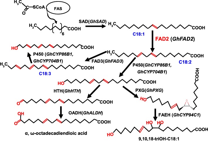
Background: In higher plants, the FAD2 gene encodes the microsomal oleate Δ12-desaturase, one of the key enzymes essential for the biosynthesis of the polyunsaturated lipids that serve many important functions in plant development and stress responses. FAD2 catalyzes the first step, in the biosynthesis of the polyunsaturated fatty acids (PUFAs) found in the cell membrane and cell wall, and it is thus of great importance to investigate the regulatory role of FAD2 in anther development.

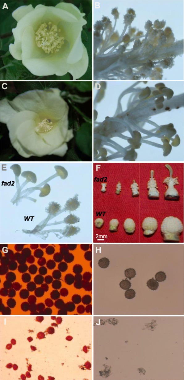
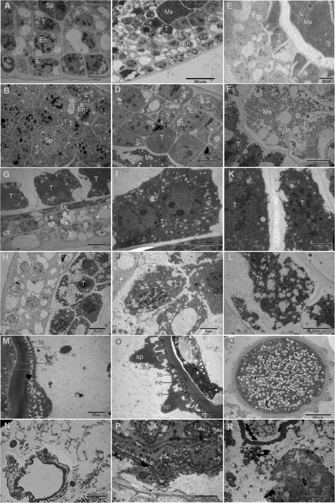
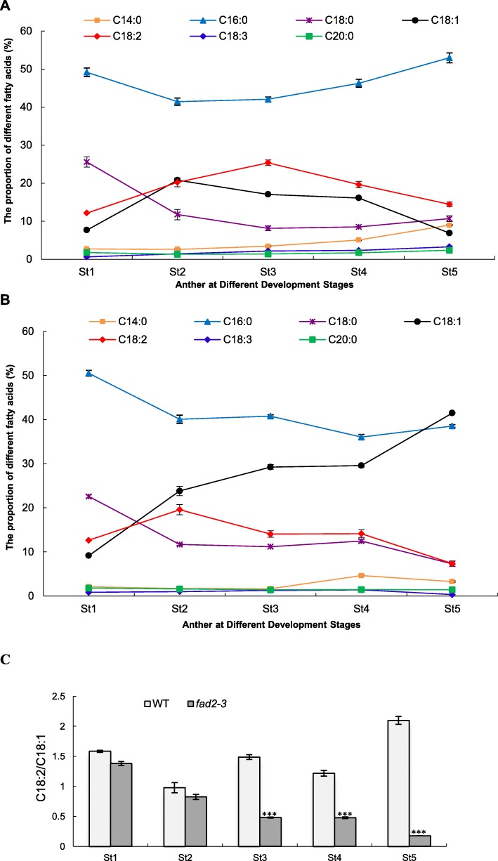
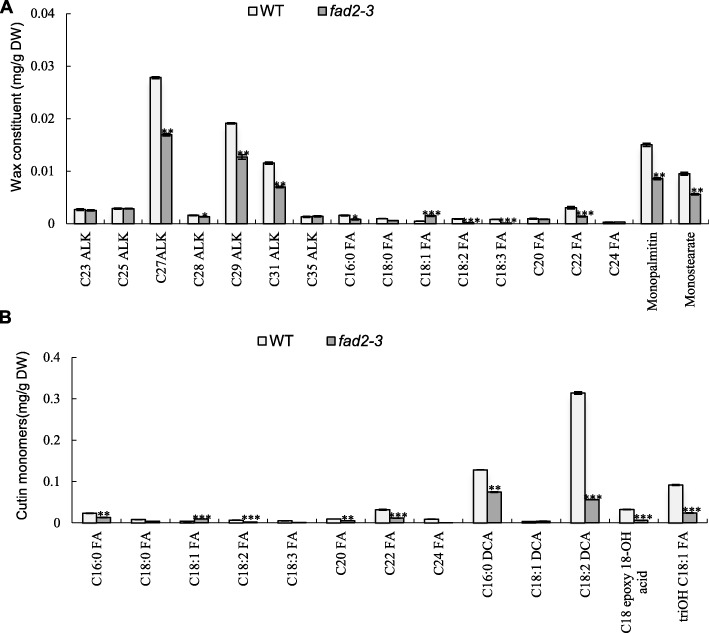
Results: We reported the molecular characterization of the cotton (Gossypium hirsutum) GhFAD2 gene family and the essential role of GhFAD2-3 in cotton anther development. G. hirsutum contains four pairs of homoeologous FAD2 genes (GhFAD2-1 to GhFAD2-4). GhFAD2-3 is ubiquitously and relatively highly expressed in all analyzed tissues, particularly in anthers. Specific inhibition of GhFAD2-3 using the RNA interference approach resulted in male sterility due to impaired anther development at the stages from meiosis to maturation. The cellular phenotypic abnormality observed at the meiosis stage of the GhFAD2-3 silenced plant (fad2-3) coincides with the significant reduction of C18:2 in anthers at the same stage. Compared with that of the wild type (WT), the content of C18:1 was 41.48%, which increased by 5 fold in the fad2-3 anther at the pollen maturation stage. Moreover, the ratio of monounsaturated to polyunsaturated fatty acid was 5.43 in fad2-3 anther, which was much higher than that of the WT (only 0.39). Through compositional analysis of anthers cuticle and transcriptome data, we demonstrated it was unfavorable to the development of anther by regulating GhFAD2-3 expression level to increase the oleic acid content.

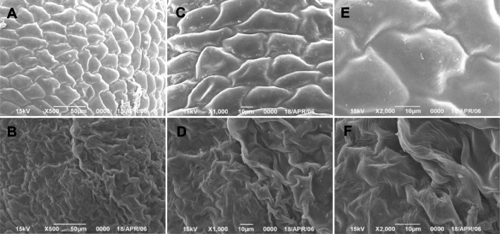
Conclusions: Our work demonstrated the importance of C18:2 and/or C18:3 in the development of the pollen exine and anther cuticle in cotton and provided clue for further investigation of the physiological significance of the fatty acid composition for plant growth and development.

Keywords: Fatty acid composition; GhFAD2; Gossypium hirsutum; Linoleic acid; Male sterile; Oleic acid.


Conflict of interest statement

The authors declare that they have no competing interests.

Figures

Fig. 1
Fig. 1
The expression profiles of GhFAD2 genes in different tissues and at different developmental stages of cotton seed and fiber. Total RNA was isolated from root, leaves, stem, anther, stigma, ovary, seed and fiber. Each sample had three biological replicates. The seeds at 40 DPA and fiber at 24 DPA were used: In seeds, the expression level of GhFAD2 reached its peak at 40 DPA [12]. It has been difficult to identify changes in gene expression for fiber after 30 DPA due to hard to extract mRNA. For each sample, a total of 3 μg RNA was used in preparing the RNA-seq library. Barcoded multiplexed RNA-seq libraries were created using the NEBNext® Ultra™ RNA Library Prep Kit for Illumina® (NEB, USA) according to the manufacturer’s protocol. Clean paired-end reads were aligned to the TM-1 reference genome, and the number of reads aligned to each gene was measured using HTSeq v0.6.1.The expression levels of individual genes were quantified using FPKM. Error bars are standard errors. Genes with an adjusted P-value < 0.05 found by DESeq were assigned as differentially expressed. * indicates a differentially expression of GhFAD2–3D between anther and other tissues (Padj < 0.05)
Fig. 2
Fig. 2
Comparison of pollen phenotype and vitality between the wild-type (WT) and the fad2–3 plants. a. Wild-type flower; b. Wild-type anthers; c. fad2–3 flower. The magnification was 5 times. d. fad2–3 anthers. The magnification was 5 times. e. Comparison of wild-type and fad2–3 mature anthers. The magnification was 5 times. f. Comparison of developing anthers from the wild-type and the fad2–3 plants; shown are de-bracted young buds collected every 4 days after bud emergence; g. Wild-type pollen grains stained by I2-KI. The magnification was 50 times. h. Wild-type pollens after acetolysis treatment. The magnification was 50 times. i. fad2–3 pollen grains stained by I2-KI. The magnification was 50 times. j. fad2–3 pollen after acetolysis treatment. The magnification was 50 times
Fig. 3
Fig. 3
Scanning electron microscopy of the mature anthers from the wild-type and the fad2–3 plants. a, c, and e. mature wild-type anthers. The magnification was 500, 1000 and 2000 times, respectively. b, d, and f. mature fad2–3 anther. The magnification was 500, 1000 and 2000 times, respectively
Fig. 4
Fig. 4
Transmission electron micrographs of anthers from the wild-type and the fad2–3 plants. A, C, E, G, I, K, M, O and Q, wild-type anthers at different developmental stages. B, D, F, H, J, L, N, P and R, fad2–3 anthers at different developmental stages. (a) Wild-type anthers with sporogenous cells and three layers of parietal cells. Bars = 5 μm. (b) fad2–3 anthers with sporogenous cells and three layers of parietal cells. Bars = 10 μm. (c) Wild-type anthers with microsporocytes and four layers of parietal cells. Bars = 20 μm. (d) fad2–3 anthers with microsporocytes and four layers of parietal cells. Bars = 5 μm. (e) Wild-type anthers at the meiosis stage. Bars = 20 μm. (f) fad2–3 anthers at the meiosis stage. Bars = 10 μm. (g) Wild-type anthers at the early tetrad stage. Bars = 5 μm. (h) fad2–3 anthers at the early tetrad stage. Bars = 10 μm. (i) High magnification of the wild-type tapetum showing double nuclei at the tetrad stage. Bars = 5 μm. (j) High magnification of the fad2–3 tapetum at the tetrad stage. Bars = 5 μm. (k) High magnification of the wild-type tapetum showing mitochondrion and endoplasmic reticulum. Bars = 2 μm. (l) High magnification of the fad2–3 tapetum showing large vacuoles, defective plastids. Bars = 2 μm. (m) Wild-type microspore at the middle developmental stage; tectum, bacula and nexine appeared. Bars = 5 μm. (n) fad2–3 microspore at the middle developmental stage; bacula and nexine appeared and showed abnormalities. Bars = 5 μm. (o) The spinules protruding from the wild-type microspore exine were formed at stages of mitosis. Bars = 5 μm. (p) fad2–3 microspore exine showing obvious abnormality. Bars = 5 μm. (q) Wild-type mature pollen grains were uniformly distributed in the small vacuole. Bars = 20 μm. (r) The degraded fad2–3 anthers exhibiting crushed cell structure. E, epidermis; En, endothecium; ML, middle layer; Sp, sporogenous cells; Ms., microsporocyte; T, tapetum; te, tectum; ne, nexine; ba, bacula; in,intine; sp., spinules
Fig. 5
Fig. 5
The proportion of fatty acid compositions in anthers at different developmental stages. a, wild-type anthers; b, fad2–3 anthers; c, the ratio of C18:2 to C18:1. Anthers at different developmental stages was used for fatty acid assay. St1-St5: Anther at different developmental stages; St1, Sporogenous cell stage; St2, Microsporocyte stage; St3, Meiosis stage; St4, Tetrad stage; St5, Pollen maturation stage. The fatty acid methyl esters were prepared by alkaline transmethylation. The analyses were performed using GCMS-QP2020 at an electron ionization of 70 eV with an HP-88 column. The quantification was carried out according to the response value of quantitative ions and the established standard curve. Each test was repeated three times, and the content of each fatty acid composition was calculated as the percentage of total measured fatty acids. The ratio of C18:2/C18:1 is calculated by dividing the relative percentage content of C18:2 from that of C18:1 at the same developmental stage. Each bar represents the mean data of three biological replicates. Error bars are standard errors. Asterisks denote significant differences to wild-type (WT) as determined by Student’s t test: ***p < 0.001
Fig. 6
Fig. 6
Analysis of anther wax and cutin in the wild type and fad2–3. (a) Wax constituents in the wild-type and fad2–3. (b) Cutin monomers in the wild-type and fad2–3. C23 ALK, tricosane; C25 ALK, pentacosane; C27 ALK, heptacosane; C28 ALK, octacosane; C29 ALK, nonacosane; C31 ALK, hentriacontane; C35 ALK, pentatriacontane. C16:0 FA, hexadecanoic acid; C18:0 FA, octadecanoic acid; C18:1 FA, 9-octadecenoic acid; C18:2 FA, 9,12-octadecadienoic acid; C18:3 FA, 9,12,15-octadecatrienoic acid; C20 FA, eicosanoic acid; C22 FA, docosanoic acid; C24 FA, tetracosanoic. C16:0 DCA, hexadecane-1,16-dioic acid; C18:1 DCA, α, ω-octadecenoic acid; C18:2 DCA, α, ω-octadecadiendioic acid; triOH C18:1 FA, 9,10,18-trihydroxy octadecenoic acid; 9,10 Epoxy 18-OH acid, 9,10-epoxy-18-OH-C18:1; DW, dry weight. The wax of anther at mature pollen stage was analyzed according to Jung et al. [26]. The wax monomer was derivatized with 1 ml BFTSA in 1 ml pyridine (1:1) for 40 min at 70 °C before GC-MS analysis. The constituent analyses were performed using GCMS-QP2020 with a DB-1 column. Each compound was quantified against the internal standard by automatic integrating the peak areas. The protocol for lipid polyester analysis was performed according to Li-Beisson et al. [50]. The cutin monomer fraction was derivatized with BFTSA/pyridine (1:1) for 60 min at 70 °C. The constituent were analyzed using GCMS-QP2020 with a DB-1 column. The GC-MS was conducted according to Li-Beisson et al. [50] with helium carrier gas at 2 ml/min. Each compound was quantified on the basis of their total ion current as described by Li-Beisson et al. [50]. Error bars are standard errors. Values represent the means ± SE, n = 3. Asterisks denote significant differences to wild-type (WT) as determined by Student’s t test: ***p < 0.001; **p < 0.01; *p < 0.05
Fig. 7
Fig. 7
qRT-PCR analysis of some candidate genes involved in the biosynthetic pathways for cutin monomers in in wild-type and fad2–3. St1-St5: Anther at different developmental stages; St1, Sporogenous cell stage; St2, Microsporocyte stage; St3, Meiosis stage; St4, Tetrad stage; St5, Pollen maturation stage. The GhFAD2–1, GhFAD3, GhSAD, GhCYP704B1, GhCYP86B1, GhHTH, GhALDH, GhPXG, and GhCYP94C1 mRNA abundance was determined by qRT-PCR, respectively. These genes encodes enzymes indicated as follows: Fatty acid desaturase 2, Fatty acid desaturase 3, Δ9-stearyl-ACP desaturase, Cytochrome P450 704B1, Cytochrome P450 86B1, ω-hydroxyacid dehydrogenase, aldehyde dehydrogenase, peroxygenase and Cytochrome P450 94C1. GhUBQ14 was used as a reference gene. All qRT–PCR reactions were performed in triplicate. Relative gene expression levels of target genes were normalized against Ct values for GhUBQ14, and the fold change (2–ΔΔCt) was determined by comparison to average expression levels. Significant differences from control were marked with * (P < 0.05), ** (P < 0.01) and *** (P < 0.001)
Fig. 8
Fig. 8
Model of FAD2 involved in the primary pathways for cutin monomers synthesis in Gossypium. Gossypium candidate genes are given in parenthesis. Arrow thickness indicates the extend of carbon flux. Enzymes presumably involved are indicated as follows: FAS, fatty acid synthase; FAD2, fatty acid desaturase 2; FAD3, fatty acid desaturase 3; SAD, stearoyl-ACP desaturase; P450, Cytochrome P450 monooxygenase; HTH, ω-hydroxyacid dehydrogenase; OADH, ω-oxo-acid dehydrogenase (aldehyde dehydrogenase); PXG, peroxygenase; FAEH, fatty acid epoxide hydrolase

Similar articles

Cited by

References

    1. Shanklin John, Cahoon Edgar B. DESATURATION AND RELATED MODIFICATIONS OF FATTY ACIDS. Annual Review of Plant Physiology and Plant Molecular Biology. 1998;49(1):611–641. - PubMed
    1. Okuley J, Lightner J, Feldmann K, Yadav N, Lark E, Browse J. Arabidopsis FAD2 gene encodes the enzyme that is essential for polyunsaturated lipid synthesis. The Plant Cell. 1994;6(1):147–158. - PMC - PubMed
    1. Jin Un-Ho, Lee Jin-Woo, Chung Young-Soo, Lee Jai-Heon, Yi Young-Byung, Kim Young-Kil, Hyung Nam-In, Pyee Jae-Ho, Chung Chung-Han. Characterization and temporal expression of a ω-6 fatty acid desaturase cDNA from sesame (Sesamum indicum L.) seeds. Plant Science. 2001;161(5):935–941.
    1. Jung Jin Hee, Kim Hyojin, Go Young Sam, Lee Saet Buyl, Hur Cheol-Goo, Kim Hyun Uk, Suh Mi Chung. Identification of functional BrFAD2-1 gene encoding microsomal delta-12 fatty acid desaturase from Brassica rapa and development of Brassica napus containing high oleic acid contents. Plant Cell Reports. 2011;30(10):1881–1892. - PubMed
    1. Li Lingyong, Wang Xiaolin, Gai Junyi, Yu Deyue. Molecular cloning and characterization of a novel microsomal oleate desaturase gene from soybean. Journal of Plant Physiology. 2007;164(11):1516–1526. - PubMed

MeSH terms

LinkOut - more resources