

OPA1: 516 unique variants and 831 patients registered in an updated centralized Variome database
- PMID: 31500643
- PMCID: PMC6734442
- DOI: 10.1186/s13023-019-1187-1
OPA1: 516 unique variants and 831 patients registered in an updated centralized Variome database
Abstract
Background: The dysfunction of OPA1, a dynamin GTPase involved in mitochondrial fusion, is responsible for a large spectrum of neurological disorders, each of which includes optic neuropathy. The database dedicated to OPA1 ( https://www.lovd.nl/OPA1 ), created in 2005, has now evolved towards a centralized and more reliable database using the Global Variome shared Leiden Open-source Variation Database (LOVD) installation.
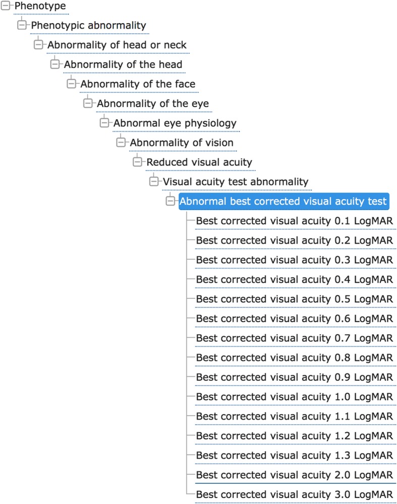
Results: The updated OPA1 database, which registers all the patients from our center as well as those reported in the literature, now covers a total of 831 patients: 697 with isolated dominant optic atrophy (DOA), 47 with DOA "plus", and 83 with asymptomatic or unclassified DOA. It comprises 516 unique OPA1 variants, of which more than 80% (414) are considered pathogenic. Full clinical data for 118 patients are documented using the Human Phenotype Ontology, a standard vocabulary for referencing phenotypic abnormalities. Contributors may now make online submissions of phenotypes related to OPA1 mutations, giving clinical and molecular descriptions together with detailed ophthalmological and neurological data, according to an international thesaurus.
Conclusions: The evolution of the OPA1 database towards the LOVD, using unified nomenclature, should ensure its interoperability with other databases and prove useful for molecular diagnoses based on gene-panel sequencing, large-scale mutation statistics, and genotype-phenotype correlations.
Keywords: Database; Dominant optic atrophy; Interoperability; Neurological disorders; OPA1; Sequence variant.
Conflict of interest statement
The authors declare that they have no competing interests.
Figures


References
-
- Kjer P. Infantile optic atrophy with dominant mode of inheritance: a clinical and genetic study of 19 Danish families. Acta Ophthalmol. 1959;164(Supp 54):1–147. - PubMed
-
- Eiberg H, Kjer B, Kjer P, Rosenberg T. Dominant optic atrophy (OPA1) mapped to chromosome 3q region. I. Linkage analysis. Hum Mol Genet. 1994;3(6):977–980. - PubMed
-
- Kjer B, Eiberg H, Kjer P, Rosenberg T. Dominant optic atrophy mapped to chromosome 3q region. II. Clinical and epidemiological aspects. Acta Ophthalmol Scand. 1996;74(1):3–7. - PubMed
Publication types
MeSH terms
Substances
LinkOut - more resources
Full Text Sources
Other Literature Sources
