. 2019 Oct 1;117(7):1342-1351.
doi: 10.1016/j.bpj.2019.07.056. Epub 2019 Aug 26.

Proton Control of Transitions in an Amino Acid Transporter

Affiliations

Proton Control of Transitions in an Amino Acid Transporter

Zhiyi Wu et al. Biophys J. .

Abstract

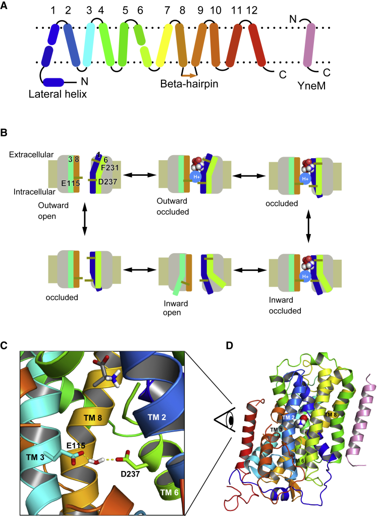
Amino acid transport into the cell is often coupled to the proton electrochemical gradient, as found in the solute carrier 36 family of proton-coupled amino acid transporters. Although no structure of a human proton-coupled amino acid transporter exists, the crystal structure of a related homolog from bacteria, GkApcT, has recently been solved in an inward-occluded state and allows an opportunity to examine how protons are coupled to amino acid transport. Our working hypothesis is that release of the amino acid substrate is facilitated by the deprotonation of a key glutamate residue (E115) located at the bottom of the binding pocket, which forms part of the intracellular gate, allowing the protein to transition from an inward-occluded to an inward-open conformation. During unbiased molecular dynamics simulations, we observed a transition from the inward-occluded state captured in the crystal structure to a much more open state, which we consider likely to be representative of the inward-open state associated with substrate release. To explore this and the role of protons in these transitions, we have used umbrella sampling to demonstrate that the transition from inward occluded to inward open is more energetically favorable when E115 is deprotonated. That E115 is likely to be protonated in the inward-occluded state and deprotonated in the inward-open state is further confirmed via the use of absolute binding free energies. Finally, we also show, via the use of absolute binding free energy calculations, that the affinity of the protein for alanine is very similar regardless of either the conformational state or the protonation of E115, presumably reflecting the fact that all the key interactions are deep within the binding cavity. Together, our results give a detailed picture of the role of protons in driving one of the major transitions in this transporter.


Figures

Figure 1
Figure 1
Structure and mechanism of GkApcT. (A) Shown is a schematic diagram of the GkApcT fold, in which the first five helices are an inverted repeat of the next five helices, similar to LeuT. The YneM helix (pink) is an additional subunit present in the crystal structure and included in the simulations presented here. (B) Mechanisms of the proton coupled transport cycle are as follows: outward-open state, outward-occluded state with substrate and proton bound, occluded state, inward-occluded state with substrate and proton ready to leave, inward-open state, and apo occluded state. (C) GkApcT in the inward-occluded state in complex with alanine is shown. The substrate alanine is drawn using the space fill representation. The two key residues gating the intracellular gate are E115 from TM 3 (aqua stick) and D237 from TM6 (green). The water molecule is not directly observed in the crystal but appears during the molecular dynamics simulations. (D) Shown is a zoomed-out view of GkApcT contextualizing the view in (C). To see this figure in color, go online.
Figure 2
Figure 2
Unbiased molecular dynamics simulations of GkApcT in a lipid bilayer. (A) Shown is a representative snapshot of the simulation system—GkApcT embedded in a POPE/POPG bilayer with alanine in the binding pocket. Sodium ions are represented as purple spheres, and chloride ions are represented as green spheres. (B) GkApcT in inward-occluded conformation and (C) inward-open conformation is shown. The conformational change from inward occluded to inward open results in an expansion of the binding cavity as represented by the white surface. In the inward-open conformation, the binding cavity extends to the bulk of intracellular space, giving rise to a potential substrate exit pathway. Some sections of helices are not displayed to help visualize the extent of the cavity. To see this figure in color, go online.
Figure 3
Figure 3
(A) Work performed as function of time in the pulling simulation. The dotted section of the line represents the state in which alanine is fully coordinated in the binding pocket. The solid section is where the interaction between the amine group of alanine and the protein is broken, whereas the interaction between the carboxyl group and protein is still intact. The dashed line represents the state in which alanine is fully dissociated from the binding pocket. Snapshots give an indication of the nature of the binding event at the various transition points. The plateaus reflect the alanine diffusing randomly within the cavity away from the binding site. (B) shows the gate in the inward-occluded state formed by A238 and V320 drawn as spheres (note no hydrogens are present in the crystal structure). (C). Shown is the path (as shown as the colored dots from red to blue as a function of time) of the alanine in an unbiased simulation that show rebinding to the binding site (indicated by the F231 residue) during the inward-open state. (D and E) show a comparison of alanine binding to the inward-occluded and inward-open states, respectively. To see this figure in color, go online.
Figure 4
Figure 4
(A and B) The space between two green spheres represents E115 and D237. The distance between the two spheres increases as GkApcT transitions from inward occluded (A) to inward open (B). This distance formed the basis of the collective variable used in later umbrella sampling calculations. (CF) Shown is the PMF calculation using umbrella sampling. The x axis is the collective variable, which is the distance between the center of mass of the Cα atoms of residues 113–117 and the center of mass of the Cα atoms of residues 235–237. The relative locations of the inward-occluded and inward-open state are annotated by red and blue arrows, respectively. PMF profiles are shown for (C) apo with deprotonated E115, (D) apo with protonated E115, (E) deprotonated with alanine in the binding site, and (F) protonated E115 with alanine bound. The width of the PMF profile represents the error associated with the potential at that point, calculated using block analysis as described in the Methods. To see this figure in color, go online.

References

    1. Krishnamurthy H., Gouaux E. X-ray structures of LeuT in substrate-free outward-open and apo inward-open states. Nature. 2012;481:469–474. - PMC - PubMed
    1. Thwaites D.T., Anderson C.M. The SLC36 family of proton-coupled amino acid transporters and their potential role in drug transport. Br. J. Pharmacol. 2011;164:1802–1816. - PMC - PubMed
    1. Wolfson R.L., Sabatini D.M. The dawn of the age of amino acid sensors for the mTORC1 pathway. Cell Metab. 2017;26:301–309. - PMC - PubMed
    1. Heublein S., Kazi S., Goberdhan D.C. Proton-assisted amino-acid transporters are conserved regulators of proliferation and amino-acid-dependent mTORC1 activation. Oncogene. 2010;29:4068–4079. - PMC - PubMed
    1. Closs E.I., Boissel J.P., Rotmann A. Structure and function of cationic amino acid transporters (CATs) J. Membr. Biol. 2006;213:67–77. - PubMed

Publication types

LinkOut - more resources