. 2019 Sep 17;117(6):1074-1084.
doi: 10.1016/j.bpj.2019.08.015. Epub 2019 Aug 22.

Positive Cooperativity in Substrate Binding by Human Thymidylate Synthase

Affiliations

Positive Cooperativity in Substrate Binding by Human Thymidylate Synthase

Jeffrey P Bonin et al. Biophys J. .

Erratum in

Abstract

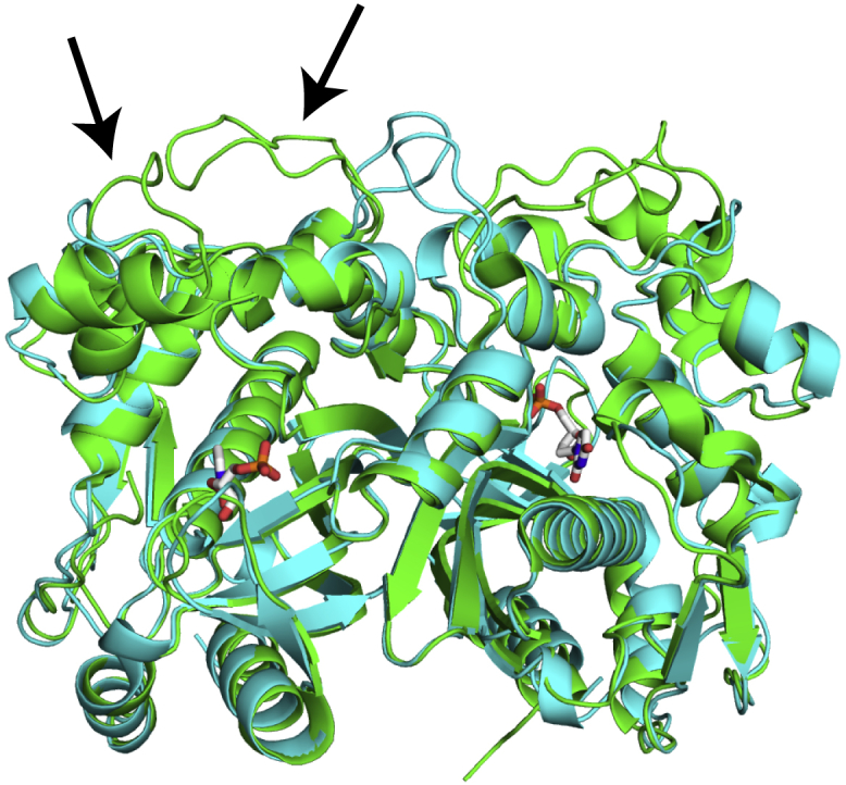
Thymidylate synthase (TS) catalyzes the production of the nucleotide dTMP from deoxyuridine monophosphate (dUMP), making the enzyme necessary for DNA replication and consequently a target for cancer therapeutics. TSs are homodimers with active sites separated by ∼30 Å. Reports of half-the-sites activity in TSs from multiple species demonstrate the presence of allosteric communication between the active sites of this enzyme. A simple explanation for the negative allosteric regulation occurring in half-the-sites activity would be that the two substrates bind with negative cooperativity. However, previous work on Escherichia coli TS revealed that dUMP substrate binds without cooperativity. To gain further insight into TS allosteric function, binding cooperativity in human TS is examined here. Isothermal titration calorimetry and two-dimensional lineshape analysis of NMR titration spectra are used to characterize the thermodynamics of dUMP binding, with a focus on quantification of cooperativity between the two substrate binding events. We find that human TS binds dUMP with ∼9-fold entropically driven positive cooperativity (ρITC = 9 ± 1, ρNMR = 7 ± 1), in contrast to the apparent strong negative cooperativity reported previously. Our work further demonstrates the necessity of globally fitting isotherms collected under various conditions, as well as accurate determination of binding competent protein concentration, for calorimetric characterization of homotropic cooperative binding. Notably, an initial curvature of the isotherm is found to be indicative of positively cooperative binding. Two-dimensional lineshape analysis NMR is also found to be an informative tool for quantifying binding cooperativity, particularly in cases in which bound intermediates yield unique resonances.


Figures

Figure 1
Figure 1
hTS and E. coli TS structures are very similar. An all-atom alignment of hTS (PDB: 5X5A, green) and E. coli TS (PDB: 1TJS, cyan) structures is shown. The only major structural differences are two loops that are extended in the human enzyme (residues 118–128, 149–156, indicated by arrows). In addition, hTS has a longer, flexible N-terminus (residues 1–25) that is not present in the crystal structure. The substrate dUMP is shown in sticks. The dUMP molecules bound to the two active sites in the enzyme are separated by ∼30 Å.
Figure 2
Figure 2
General two-site binding model. The binding polynomial, P, is used to determine the populations of bound species (apo, lig1, lig2) throughout titration based on the two macroscopic binding affinities for both ITC and NMR studies presented here. Note that this model fixes the stoichiometry at n = 2, which is justified by the x-ray model (Fig. 1), as well as our NMR titration spectra (Fig. 5). This model does not presume cooperative or independent binding. Rather, separate macroscopic affinities are fitted, and the presence or absence of cooperativity is assessed afterwards. In the case of independent binding, the two microscopic affinities would be identical because of the homodimeric nature of the enzyme. On the other hand, cooperativity in the binding would introduce a difference in the values of the two microscopic affinities, as discussed in the text. In addition to the two macroscopic affinities and enthalpies, our ITC fits include a parameter that scales the given protein concentration, accounting for error in the predicted extinction coefficient and/or presence of binding incompetent fraction. This parameter is not fitted for the NMR titration, but rather, a fixed scaling factor of 0.75 is used based on our PULCON-NMR result.
Figure 3
Figure 3
hTS binds dUMP with positive cooperativity. (a) Global fit of ITC isotherms collected at various hTS concentrations in 20 mM NaCl at 25°C is shown. The low-mole-ratio points in green are designed to determine the first enthalpy, which shows a correlation with the first affinity in some of our fits (Fig. S1). Boxed points highlight the distinct initial curvature present in our high c-value data, which we find to be indicative of positive cooperativity. (b) Thermodynamic parameters from 150 Monte Carlo simulated data sets showing changes in enthalpy for the two dUMP binding events (red bars) and similarly for the corresponding two entropic contributions (blue bars) are given. A ∼9-fold increase in the second affinity relative to the first (black bars) is observed, and this is driven by the >4 kcal/mol more negative entropic contribution for the second binding event. A complete table of parameters is found in Table S1. (c) Simulation of isotherms at 400 μM protein concentration using the fitted enthalpies shows the presence of a large initial curvature only in the case of positive cooperativity. This curvature is clearly seen in 246 μM data in Fig. 3a. For each curve, the fitted first macroscopic affinity is used and the second macroscopic affinity is adjusted to give the indicated cooperativity. (d) The reason for this initial curvature is seen by looking at the change in the equilibrium populations with each injection (Δlig1 and Δlig2) during titration; Δlig1 has a large negative slope at low mole ratio. Although there is also a large positive slope in Δlig2, the slope of Δlig1 is greater in magnitude, leading to the large initial curvature seen in the Δlig1 + Δlig2 curve. Note that the isotherms are simply this Δlig1 + Δlig2 curve with Δlig1 weighted by ΔH1 and Δlig2 weighted by ΔH1 + ΔH2.
Figure 4
Figure 4
Temperature dependence of dUMP binding thermodynamics. Global fit of binding isotherms at temperatures ranging from 5 to 35°C using van’t Hoff temperature dependence of the macroscopic affinities is shown (top). The low-mole-ratio points in yellow are designed to determine the first enthalpy, which shows a correlation with the first affinity in some of our fits (Fig. S1). A bar graph of cooperative thermodynamic parameters from 150 Monte Carlo simulated data sets (bottom) shows positive cooperativity in dUMP binding across this temperature range. Cooperative parameters are defined as the difference between these parameters for the two binding events (Δh = ΔH2ΔH1, etc.). Note that Δgmicro = −RTln(ρ). The observed Δgmicro values of ∼−1 kcal/mol correspond to ρ ∼ 5. The large jump in Δh (and TΔsmicro) from 25 to 35°C results from a significant change in the binding heat capacities in this range. This is consistent with an equilibrium coupled to the binding, as described in the text. A complete table of parameters is found in Table S2.
Figure 5
Figure 5
Positive binding cooperativity determined by NMR titration. (a) Quartet resonance pattern for Asp48 of hTS at 1:1 ratio of substrate/dimer, as seen in our titration, is shown. The presence of distinct resonances for the singly bound state makes these NMR signals well suited for determination of macroscopic affinities for multiple binding events. (b) Fit of 170 μM (after application of 0.75 scaling factor) hTS dimer titration spectra for Asp48 using TITAN software is shown. For each titration point, three-dimensional plots (left) as well as 2D contour plots (right) of the Asp48 resonances from our data (blue) overlaid with reconstructed resonances from our fit parameters (red) are shown. The number of resonances present at each mole ratio, as well as their linewidths and intensities, are well-captured by our fit. Other spin systems used in the global fit can be seen in Fig. S6. (c) A box plot of ρ-values obtained from 150 Monte Carlo simulated ITC data sets and 100 bootstrap resampled NMR titration spectra shows excellent agreement in the relative microscopic affinities between the two approaches. A table of parameters for the NMR titration fit is found in Table S3.

References

    1. Cui Q., Karplus M. Allostery and cooperativity revisited. Protein Sci. 2008;17:1295–1307. - PMC - PubMed
    1. Monod J., Wyman J., Changeux J.P. On the nature of allosteric transitions: a plausible model. J. Mol. Biol. 1965;12:88–118. - PubMed
    1. Motlagh H.N., Wrabl J.O., Hilser V.J. The ensemble nature of allostery. Nature. 2014;508:331–339. - PMC - PubMed
    1. Guo J., Zhou H.X. Protein allostery and conformational dynamics. Chem. Rev. 2016;116:6503–6515. - PMC - PubMed
    1. Lisi G.P., Loria J.P. Solution NMR spectroscopy for the study of enzyme allostery. Chem. Rev. 2016;116:6323–6369. - PMC - PubMed

Publication types

LinkOut - more resources