Review
. 2019 Sep;27S(Suppl):S104-S113.
doi: 10.1016/j.molmet.2019.06.013.

β cell responses to inflammation

Affiliations
Review

β cell responses to inflammation

Sahar Usmani-Brown et al. Mol Metab. 2019 Sep.

Abstract

Background: The extended and clinically silent progression of Type 1 diabetes (T1D) creates a challenge for clinical interventions and for understanding the mechanisms that underlie its pathogenesis. Over the course of the development of Type 1 diabetes, studies in animal models and of human tissues have identified adaptive changes in β cells that may affect their immunogenicity and susceptibility to killing. Loss of β cells has traditionally been identified by impairment in function but environmental factors may affect these measurements.

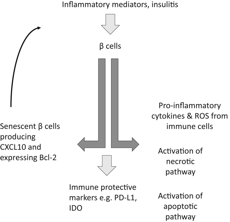
Scope of review: In this review we will highlight features of β cell responses to cell death, particularly in the setting of inflammation, and focus on methods of detecting β cell death in vivo.

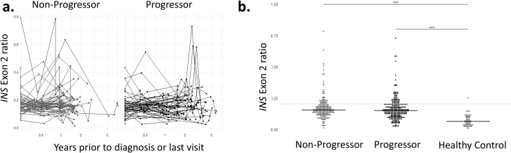
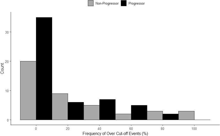
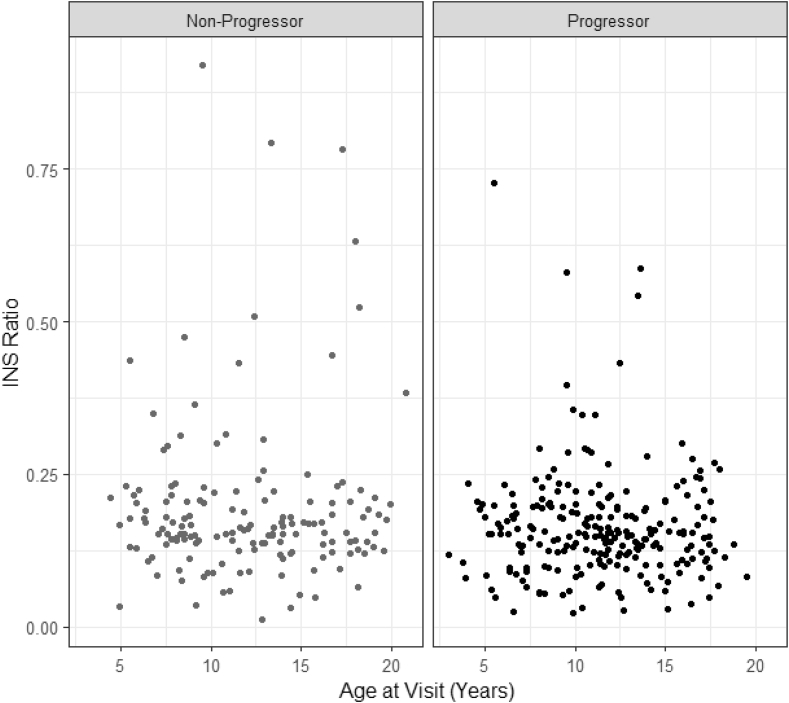
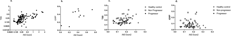
Major conclusions: We developed an assay to measure β cell death in vivo by detecting cell free DNA with epigenetic modifications of the INS gene that are found in β cells. This assay has robust technical performance and identifies killing in individuals at very high risk for disease, but its ability to identify β cell killing in at-risk relatives is limited by the short half-life of the cell free DNA and the need for repeated sampling over an extended course. We present results from the Diabetes Prevention Trial-1 using this assay. In addition, recent studies have identified cellular adaptations in some β cells that may avoid killing but impair metabolic function. Cells with these characteristics may aggravate the autoimmune response but also may represent a potentially recoverable source of functional β cells.

Keywords: Apoptosis; Beta cells; Death; Inflammation; Methylation; Type 1 diabetes.


Figures

Figure 1
Figure 1
β cell death and survival. Recent studies of the fate of β cells during the progression of autoimmune diabetes in animal models and humans suggest that there may be alternate fates of cells that may lead to destruction, survival, or even acceleration of the immune response. Inflammatory mediators may induce changes in β cells that may lead to their demise through classical pathways such as Fas/FasL, cytolysis, or other mechanisms. However, there are also adaptive changes in β cells that may protect them from further destruction, such as increased expression of PD-L1 and IDO, or may also enhance the autoimmune response by the development of senenscent cells that can produce chemokines (e.g. CXCL10) that can recruit immune cells, or inflammatory cytokines (e.g. IL-6) that have direct toxic effects. Thus, there is a dynamic response to inflammatory stressors that may ultimately determine whether cells are killed by autoimmune responses or protected.
Figure 2
Figure 2
Ratios of unmethylated to total (unmethylated + methylated) INS DNA in the Diabetes Prevention Trial-1. The ratios of unmethylated:total INS DNA are shown over time in DPT-1 participants who did (progressors) or did not (non-progressors) develop T1D during the trial. (a) The time represents the years prior to the clinical diagnosis of T1D or the last study visit. Each line shows, with symbols, the measurements for an individual over time. (b) all of the data points from the progressors (n = 55) and non-progressors (n = 42) are shown. The levels were significantly higher in the progressors and non-progressors compared to healthy control subjects (***p < 0.001, ANOVA) but not to each other.
Figure 3
Figure 3
Elevated unmethylated INS ratios in the DPT-1 trial. The number of elevated (>0.203) levels are shown for all of the data shown in Figure 1B for the progressors (n = 97) and non-progressors (n = 42). The threshold of 0.203 represents the 95th percentile of the ratios from healthy control subjects (n = 165).
Figure 4
Figure 4
Relationship between age and the INS ratio in the DPT-1 in children. The relationships between the ratio of unmeth:total INS DNA is shown by age of the participants. The plot shows 504 individual observations (progressors = 310, non-progressors = 194).
Figure 5
Figure 5
Relationship between measures of unmethylated INS DNA. (a,b) a comparison of the unmethylated:total (unmethylated + methylated) INS DNA detected with (a) the Exon2 or the transcription start site (TSS) probe or the (b) Exon 2 and IGRP probe are shown in patients who received islet autotransplants. In (a) all of the samples (127 pairs) over the first 3 days are shown (r = 0.66, p < 0.0001, Pearson) and (b) a subset (10 pairs) of the samples (r = 0.69, p = 0.027, Pearson) (c) The relationship between the Exon 2 (r = 0.34, p = 0.005, Pearson)and TSS probe and (d) the Exon 2 and IGRP probe (r = −.01, p = ns) in a subset of samples from DPT-1 participants (n = 66 pairs).

References

    1. Bluestone J.A., Herold K., Eisenbarth G. Genetics, pathogenesis and clinical interventions in type 1 diabetes. Nature. 2010;464:1293–1300. - PMC - PubMed
    1. Herold K., Vignali D.A., Cooke A., Bluestone J. Type 1 diabetes: translating mechanistic observations into effective clinical outcomes. Nature Reviews Immunology. 2013;13(4):243–256. - PMC - PubMed
    1. Keenan H.A., Sun J.K., Levine J., Doria A., Aiello L.P., Eisenbarth G. Residual insulin production and pancreatic ss-cell turnover after 50 years of diabetes: Joslin Medalist Study. Diabetes. 2010;59:2846–2853. - PMC - PubMed
    1. Oram R.A., Jones A.G., Besser R.E., Knight B.A., Shields B.M., Brown R.J. The majority of patients with long-duration type 1 diabetes are insulin microsecretors and have functioning beta cells. Diabetologia. 2014;57:187–191. - PMC - PubMed
    1. Professional practice committee for the standards of medical care in diabetes-2016. Diabetes Care. 2016;39(Suppl 1):S107–S108. - PubMed

Publication types