Review
. 2020 Apr 1;10(4):a037424.
doi: 10.1101/cshperspect.a037424.

The Immune Microenvironment and Cancer Metastasis

Affiliations
Review

The Immune Microenvironment and Cancer Metastasis

Asmaa El-Kenawi et al. Cold Spring Harb Perspect Med. .

Abstract

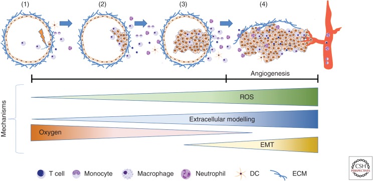
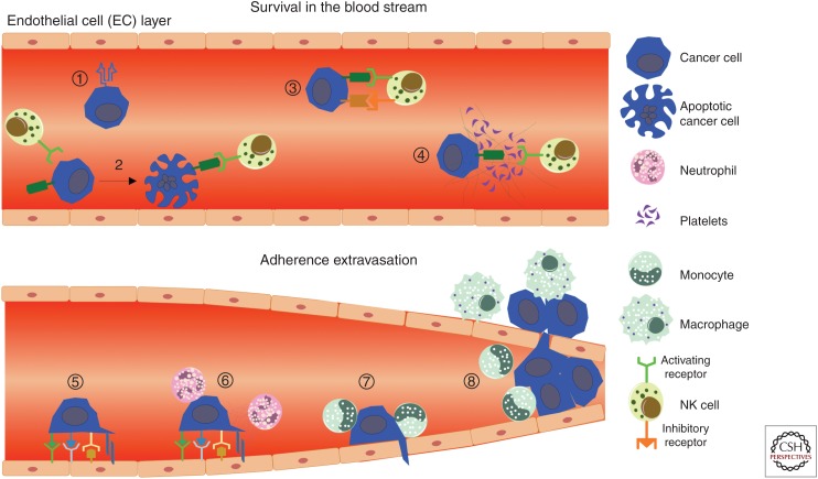
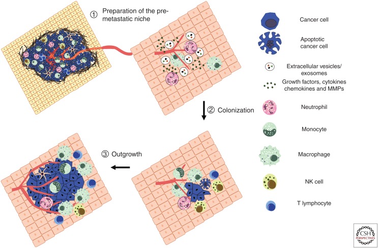
The dynamic interplay between neoplastic cells and the immune microenvironment regulates every step of the metastatic process. Immune cells contribute to invasion by secreting a cornucopia of inflammatory factors that promote epithelial-to-mesenchymal transition and remodeling of the stroma. Cancer cells then intravasate to the circulatory system assisted by macrophages and use several pathways to avoid recognition by cytotoxtic lymphocytes and phagocytes. Circulating tumor cells that manage to adhere to the vasculature and encounter premetastic niches are able to use the associated myeloid cells to extravasate into ectopic organs and establish a dormant microscopic colony. If successful at avoiding repetitive immune attack, dormant cells can subsequently grow into overt, clinically detectable metastatic lesions, which ultimately account to most cancer-related deaths. Understanding how disseminated tumor cells evade and corrupt the immune system during the final stages of metastasis will be pivotal in developing new therapeutic modalities that combat metastasis.


Figures

Figure 1.
Figure 1.
Establishment of the tumor immune microenvironment: Environmental insults or spontaneous genetic alterations in epithelial cells can lead to immune surveillance (1) or the growth of premalignant cells that evade clearance by innate and adaptive immune cells (2). In addition to driving cell proliferation, certain oncogenic alterations are associated with the release of cytokines that recruit additional immune cells to aid neoplastic progression by providing mitogenic factors or producing reactive oxygen species that result in the accumulation of additional genetic mutations (3). Uncontrolled cellular growth at this stage causes environmental changes such as reduced oxygen levels (hypoxia), extracellular matrix (ECM) turnover, and stromal disruption (4). These signals of tissue damage result in a pathological healing response that includes the creation of a disordered and permeable vascular network (angiogenic switch), as well as morphological and transcriptional changes in epithelial cells that reflect a mesenchymal phenotype (EMT). Malignant cells are thus able to migrate toward and intravasate into the vascular lumen, becoming circulating tumor cells.
Figure 2.
Figure 2.
Immune evasion and assistance during cancer cell dissemination. During circulation within the intravascular space, major histocompatibility complex (MHC) I expression protects by circulating tumor cells (CTCs) from natural killer (NK) cell recognition (1), whereas down-regulation of MHC I and up-regulation of NK cell activating ligands can lead to cytotoxicity (2). Conversely, recognition can be antagonized via up-regulation of NK cell inhibitory ligands (3) or physical shielding using platelet/fibrin coagulates (4). To extravasate into the tissue, CTCs interact with endothelial cells via selectins, cadherins, integrins, CD44, junctional adhesion molecules (5). Neutrophils are able to assist this process via ICAM-1 and VCAM-1 interactions, as well as through the release of NETs that trap CTCs and hide the cells from lympohcytes (6). Recruitment of CCR2+Ly6C+ monocytes (7) results in local production of VEGF, thereby increasing vascular permeability to facilitate CTC extravasation (8).
Figure 3.
Figure 3.
Establishment of the metastatic immune microenvironment: The primary tumor can actively prime organs by forming a metastasis-permissive microenvironment before the arrival of disseminated tumor cells (DTCs) (1). This premetastatic niche includes recruited monocytes, macrophages, and neutrophils, as well as tumor-secreted factors such as cytokines, chemokines, growth factors, and proteases. Enhanced vascular leakiness, reorganization of the stroma and extracellular matrix, and the establishment of an immunosuppressive environment are hallmarks of the metastatic microenvironment (2). These allow DTCs to evade natural killer (NK) cell immunity and colonize the ectopic organ as single cells or micrometastatic lesions. Micrometastatic lesions often enter a period of dormancy, as they are ill-adapted to the new microenvironment, encounter growth suppressive factors such as TGF-β, and are subjected to NK cell-mediated immune surveillance. Outgrowth involves tumor-intrinsic changes alongside the induction of angiogenesis and the creation of a mature immunosuppressive microenvironment to evade recognition by cytotoxic lymphocytes (3).

References

    1. Abba MC, Gong T, Lu Y, Lee J, Zhong Y, Lacunza E, Butti M, Takata Y, Gaddis S, Shen J, et al. 2015. A molecular portrait of high-grade ductal carcinoma in situ. Cancer Res 75: 3980–3990. 10.1158/0008-5472.CAN-15-0506 - DOI - PMC - PubMed
    1. Adams GN, Rosenfeldt L, Frederick M, Miller W, Waltz D, Kombrinck K, McElhinney KE, Flick MJ, Monia BP, Revenko AS, et al. 2015. Colon cancer growth and dissemination relies upon thrombin, stromal PAR-1, and fibrinogen. Cancer Res 75: 4235–4243. 10.1158/0008-5472.CAN-15-0964 - DOI - PMC - PubMed
    1. Aird WC. 2007. Phenotypic heterogeneity of the endothelium: I. Structure, function, and mechanisms. Circ Res 100: 158–173. 10.1161/01.RES.0000255691.76142.4a - DOI - PubMed
    1. Akkari L, Gocheva V, Kester JC, Hunter KE, Quick ML, Sevenich L, Wang HW, Peters C, Tang LH, Klimstra DS, et al. 2014. Distinct functions of macrophage-derived and cancer cell-derived cathepsin Z combine to promote tumor malignancy via interactions with the extracellular matrix. Genes Dev 28: 2134–2150. 10.1101/gad.249599.114 - DOI - PMC - PubMed
    1. Albrengues J, Shields MA, Ng D, Park CG, Ambrico A, Poindexter ME, Upadhyay P, Uyeminami DL, Pommier A, Kuttner V, et al. 2018. Neutrophil extracellular traps produced during inflammation awaken dormant cancer cells in mice. Science 361: eaao4227 10.1126/science.aao4227 - DOI - PMC - PubMed

Publication types

MeSH terms

LinkOut - more resources