

Hepatitis C Virus Structure: Defined by What It Is Not
- PMID: 31501263
- PMCID: PMC6938659
- DOI: 10.1101/cshperspect.a036822
Hepatitis C Virus Structure: Defined by What It Is Not
Abstract
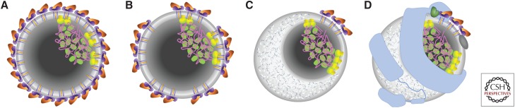
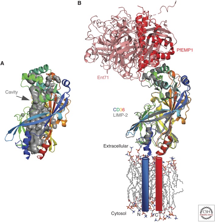
Hepatitis C virus (HCV) represents an important and growing public health problem, chronically infecting an estimated 70 million people worldwide. This blood-borne pathogen is generating a new wave of infections in the United States, associated with increasing intravenous drug use over the last decade. In most cases, HCV establishes a chronic infection, sometimes causing cirrhosis, end-stage liver disease, and hepatocellular carcinoma. Although a curative therapy exists, it is extremely expensive and provides no barrier to reinfection; therefore, a vaccine is urgently needed. The virion is asymmetric and heterogeneous with the buoyancy and protein content similar to low-density lipoparticles. Core protein is unstructured, and of the two envelope glycoproteins, E1 and E2, the function of E1 remains enigmatic. E2 is responsible for specifically binding host receptors CD81 and scavenger receptor class B type I (SR-BI). This review will focus on structural progress on HCV virion, core protein, envelope glycoproteins, and specific host receptors.
Copyright © 2020 Cold Spring Harbor Laboratory Press; all rights reserved.
Figures


Similar articles
-
Hepatitis C virus E2 envelope glycoprotein core structure.Science. 2013 Nov 29;342(6162):1090-4. doi: 10.1126/science.1243876. Science. 2013. PMID: 24288331 Free PMC article.
-
Conformational Flexibility in the Immunoglobulin-Like Domain of the Hepatitis C Virus Glycoprotein E2.mBio. 2017 May 16;8(3):e00382-17. doi: 10.1128/mBio.00382-17. mBio. 2017. PMID: 28512091 Free PMC article.
-
A Recombinant Hepatitis C Virus Genotype 1a E1/E2 Envelope Glycoprotein Vaccine Elicits Antibodies That Differentially Neutralize Closely Related 2a Strains through Interactions of the N-Terminal Hypervariable Region 1 of E2 with Scavenger Receptor B1.J Virol. 2019 Oct 29;93(22):e00810-19. doi: 10.1128/JVI.00810-19. Print 2019 Nov 15. J Virol. 2019. PMID: 31462563 Free PMC article.
-
Hypervariable Region 1 in Envelope Protein 2 of Hepatitis C Virus: A Linchpin in Neutralizing Antibody Evasion and Viral Entry.Front Immunol. 2018 Sep 27;9:2146. doi: 10.3389/fimmu.2018.02146. eCollection 2018. Front Immunol. 2018. PMID: 30319614 Free PMC article. Review.
-
Hepatitis C Virus Envelope Glycoproteins: A Balancing Act of Order and Disorder.Front Immunol. 2018 Aug 24;9:1917. doi: 10.3389/fimmu.2018.01917. eCollection 2018. Front Immunol. 2018. PMID: 30197646 Free PMC article. Review.
Cited by
-
An alternate conformation of HCV E2 neutralizing face as an additional vaccine target.Sci Adv. 2020 Jul 24;6(30):eabb5642. doi: 10.1126/sciadv.abb5642. eCollection 2020 Jul. Sci Adv. 2020. PMID: 32754640 Free PMC article.
-
The Malignant Transformation of Viral Hepatitis to Hepatocellular Carcinoma: Mechanisms and Interventions.MedComm (2020). 2025 Mar 8;6(3):e70121. doi: 10.1002/mco2.70121. eCollection 2025 Mar. MedComm (2020). 2025. PMID: 40060195 Free PMC article. Review.
-
Viral oncogenesis in cancer: from mechanisms to therapeutics.Signal Transduct Target Ther. 2025 May 12;10(1):151. doi: 10.1038/s41392-025-02197-9. Signal Transduct Target Ther. 2025. PMID: 40350456 Free PMC article. Review.
-
Understanding the relationship between HCV infection and progression of kidney disease.Front Microbiol. 2024 Jun 28;15:1418301. doi: 10.3389/fmicb.2024.1418301. eCollection 2024. Front Microbiol. 2024. PMID: 39006752 Free PMC article. Review.
-
Cellular Organelles Involved in Hepatitis E Virus Infection.Pathogens. 2021 Sep 17;10(9):1206. doi: 10.3390/pathogens10091206. Pathogens. 2021. PMID: 34578238 Free PMC article. Review.
References
-
- Banda DH, Perin PM, Brown RJP, Todt D, Solodenko W, Hoffmeyer P, Kumar Sahu K, Houghton M, Meuleman P, Muller R, et al. 2019. A central hydrophobic E1 region controls the pH range of hepatitis C virus membrane fusion and susceptibility to fusion inhibitors. J Hepatol 70: 1082–1092. 10.1016/j.jhep.2019.01.033 - DOI - PubMed
-
- Bankwitz D, Steinmann E, Bitzegeio J, Ciesek S, Friesland M, Herrmann E, Zeisel MB, Baumert TF, Keck ZY, Foung SK, et al. 2010. Hepatitis C virus hypervariable region 1 modulates receptor interactions, conceals the CD81 binding site, and protects conserved neutralizing epitopes. J Virol 84: 5751–5763. 10.1128/JVI.02200-09 - DOI - PMC - PubMed
-
- Bartosch B, Vitelli A, Granier C, Goujon C, Dubuisson J, Pascale S, Scarselli E, Cortese R, Nicosia A, Cosset FL. 2003. Cell entry of hepatitis C virus requires a set of co-receptors that include the CD81 tetraspanin and the SR-B1 scavenger receptor. J Biol Chem 278: 41624–41630. 10.1074/jbc.M305289200 - DOI - PubMed
Publication types
MeSH terms
Substances
LinkOut - more resources
Full Text Sources
Research Materials