. 2019 Nov 5;201(23):e00365-19.
doi: 10.1128/JB.00365-19. Print 2019 Dec 1.

Chromosomal Recombination Targets in Chlamydia Interspecies Lateral Gene Transfer

Affiliations

Chromosomal Recombination Targets in Chlamydia Interspecies Lateral Gene Transfer

Robert J Suchland et al. J Bacteriol. .

Abstract

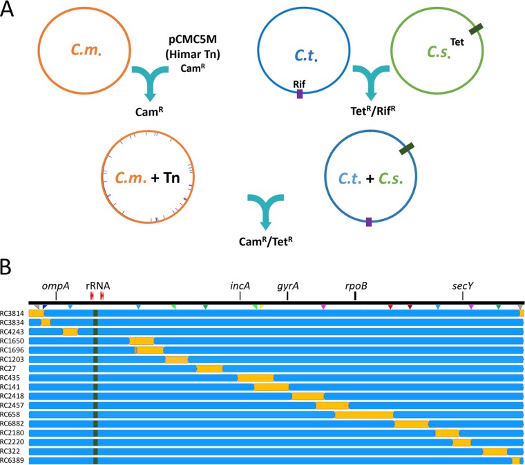
Lateral gene transfer (LGT) among Chlamydia trachomatis strains is common, in both isolates generated in the laboratory and those examined directly from patients. In contrast, there are very few examples of recent acquisition of DNA by any Chlamydia spp. from any other species. Interspecies LGT in this system was analyzed using crosses of tetracycline (Tc)-resistant C. trachomatis L2/434 and chloramphenicol (Cam)-resistant C. muridarum VR-123. Parental C. muridarum strains were created using a plasmid-based Himar transposition system, which led to integration of the Camr marker randomly across the chromosome. Fragments encompassing 79% of the C. muridarum chromosome were introduced into a C. trachomatis background, with the total coverage contained on 142 independent recombinant clones. Genome sequence analysis of progeny strains identified candidate recombination hot spots, a property not consistent with in vitroC. trachomatis × C. trachomatis (intraspecies) crosses. In both interspecies and intraspecies crosses, there were examples of duplications, mosaic recombination endpoints, and recombined sequences that were not linked to the selection marker. Quantitative analysis of the distribution and constitution of inserted sequences indicated that there are different constraints on interspecies LGT than on intraspecies crosses. These constraints may help explain why there is so little evidence of interspecies genetic exchange in this system, which is in contrast to very widespread intraspecies exchange in C. trachomatisIMPORTANCE Genome sequence analysis has demonstrated that there is widespread lateral gene transfer among strains within the species C. trachomatis and with other closely related Chlamydia species in laboratory experiments. This is in contrast to the complete absence of foreign DNA in the genomes of sequenced clinical C. trachomatis strains. There is no understanding of any mechanisms of genetic transfer in this important group of pathogens. In this report, we demonstrate that interspecies genetic exchange can occur but that the nature of the fragments exchanged is different than those observed in intraspecies crosses. We also generated a large hybrid strain library that can be exploited to examine important aspects of chlamydial disease.

Keywords: Chlamydia; lateral gene transfer; recombination; transposon.


Figures

FIG 1
FIG 1
Generating interspecies recombinants from crosses between C. muridarum and C. trachomatis parents. (A) Transposon mutagenesis, as described in reference , led to the creation of a variety of randomly mutated, chloramphenicol-resistant C. muridarum strains (C.m. + Tn). C. trachomatis parents were generated by transferring the C. suis tet(C) locus, plus adjacent sequences, to rifampin (Rif)-resistant C. trachomatis strains D/UW3, L2/434, and F/70, using previously described methods (8). Cocultures of individual C. muridarum and C. trachomatis parent strains were then incubated in the presence of both Cam and Tc to select for recombinant progeny. (B) Doubly antibiotic-resistant strains were cloned by limiting dilution, and their genomes were sequenced. A linear representation of the C. trachomatis chromosome is shown (black line). The locations of selected genes are included for reference purposes. The location of the tet(C) locus is indicated with green shading in the individual recombinants and is in this position in each of the progeny strains generated by LGT. Seventeen individual recombinant strains (orange bars), which collectively represent 79% of the C. muridarum genome, are indicated. The complete list of recombinant strains is given in Fig. S1 and Table S1.
FIG 2
FIG 2
Recombinant progeny generated from mixed cultures of C. muridarum (Camr) and C. trachomatis (Tcr). Each black bar represents a segment of C. muridarum DNA transferred into an individual C. trachomatis parent. The three grey bars are clones generated without the Camr transposon insert. All the strains shown are cloned recombinant progeny from individual LGT experiments. The inset shows an expanded view of the chromosomal region containing the rRNA operons (red). The green sequences within the inset represent reciprocal clones, in which C. trachomatis L2tet9 DNA transferred into various C. muridarum backgrounds. The indicated reciprocal clone (RC1361) is described in Fig. S1 and Table S1. The orange bars show different sets of recombinants that are compared in Fig. 6. The transposon insertion sites of the individual parents used are shown as colored triangles and are indicated with tick marks in the relevant recombinant strains. Gene identities are included as in Fig. 1. The red target symbol near the center of the genome is the chromosomal ter sequence, a recombination hot spot. For scale, the rRNA operons and intervening sequence equal 43 kb.
FIG 3
FIG 3
Recombination margins in Chlamydia intraspecies and interspecies lateral gene transfer events. The percentages of identity at the collected margins of recombination for chlamydial interspecies (A) and intraspecies (B) events are indicated for the collected primary recombination events. Intraspecies data were collected from the recombinant strains described by Jeffrey et al. (5). In each graph, the x axis indicates the percent identity between the parental strains of the recombinant progeny, and the y axis depicts the number of recombination margins falling in the indicated range. The gray bars indicate the percent identity within the 100 bp surrounding a crossover site (50 per side), and the black bars indicate the percent identity within the 1,000 bp surrounding a crossover site. The dashed line in each graph indicates the overall percent genomic identity for parents in the specific crosses.
FIG 4
FIG 4
Examination of recombinant clones that express a growth defect. (A) Inclusion-forming units were determined at three time points following inoculation of McCoy cells with a set of four recombinants plus the C. trachomatis L2tet9 parent. The numbers on the vertical axis are inclusion-forming units per milliliter of lysed, infected cells. Monolayers were inoculated with approximately 30,000 IFU per ml. The error bars represent standard deviations. (B) Map of the recombinants tested in the growth experiments. This is a subset of the library of recombinants shown in Fig. 2 and includes recombinants from regions D (RC27) and E (RC435, RC745, and RC1219) in Fig. 2. The locations of incA and the 32-bp recombination hot spot can be identified here and in Fig. 2 for positioning. The triangle indicates the homologous position in C. muridarum that is targeted in transposon mutant CM013, for which no syntenic recombinants were generated. For scale, the C. muridarum insert shown for recombinant RC27 is 52 kb. (C) Immunofluorescence microscopic images of a methanol-fixed McCoy cell recombinant progeny strain with a wild-type inclusion morphology phenotype (RC27) and a recombinant expressing the early-lysis phenotype (RC745). The cells were fixed at 30 h and had identical multiplicities of infection. Red, MOMP; blue, DNA.
FIG 5
FIG 5
Examination of progeny strains using nonhomologous target sites in recombination. Strains RC936 (A) and RC1201 (B and C) have sequences that result from one (RC1201) or both (RC936) recombination endpoints occurring at nonhomologous target sites. (A and B) C. muridarum sequence is indicated in red, while C. trachomatis sequences are indicated in blue or black. In both cases, the predicted target of recombination shares no immediate identity with the C. muridarum donor DNA. The actual site of recombination, indicated with an asterisk above each linear chromosome, is either identical with the donor DNA sequence (RC1201) or very nearly identical (RC936). The tables show the numbers of occurrences in the recipient chromosome for increasingly long candidate target sites. In panel A, a second, identical target sequence is identified within rpoN. This target was not used in any identified recombinant. (C) The illegitimate recombination event at one end of the integrated DNA led to a duplication of ∼16 kb of chlamydial DNA. The recipient DNA (C. trachomatis; blue, with hatched ORFs) and the donor DNA (C. muridarum; tan, with solid ORFs) are largely homologous in this region. This clone carries homologous copies of incA (red) from each species, and the protein products of both genes are expressed and correctly localized to the inclusion membrane (green labeling in the micrographs). Host cell nuclei are labeled blue with DAPI (4′,6-diamidino-2-phenylindole) in each micrograph.
FIG 6
FIG 6
Lengths of primary recombined fragments generated in C. muridarum × C. trachomatis crosses. (A) Recombinant fragment lengths are indicated on the vertical axis, and the groups from Fig. 2 are listed on the horizontal axis. Each region shown in Fig. 2 that is represented by at least 6 individual clones was included in these analyses. The number of clones examined in each region is indicated in parentheses. The overall mean from all primary C. muridarum inserts into the C. trachomatis genome is indicated as a dashed line, while group medians are shown with solid lines within the violin plots. (B) Parallel data from recombinant progeny generated in intraspecies (C. trachomatis × C. trachomatis) recombinant strains described in reference 5. Note the different scales on the vertical axes in panels A and B. The difference between the mean length for each region and the overall mean for all recombined inserts was analyzed using a Weibull distribution. *, P value of ∼0.05; **, P < 0.05; ***, P < 0.001; n.s., not significant.
FIG 7
FIG 7
Open reading frame map of the plasticity zones for parental strains C. trachomatis L2tet9, C. muridarum transposon mutant CM007, and three PZ-based recombinants strains (KU2043, RC768, and RC215). C. muridarum ORFs are indicated with solid colors, while C. trachomatis ORFs are indicated with hatched colors. Homologous ORFs between strains are shown in black, green, orange, and blue. Plasticity zone genes unique to each species are shown in light gray, and the C. muridarum tox loci (truncated) are yellow. The transposed sequence within macP (TC0431) contributed by the CM007 parent is indicated in red. The 32-bp recombination target (ter) is indicated with a bullseye symbol. Homologous recombination at this target sequence forms a crossover point in each of the PZ-containing recombinant strains. Gene numbers are indicated by the species of origin.
FIG 8
FIG 8
The 32-bp recombination target within the chlamydial PZ is conserved among strains and forms the chlamydial replication termination sequence. (A) BLAST analysis of the 32-bp recombination target among Chlamydia spp., Chlamydia-like organisms, and E. coli. The genomic sources of these data are indicated in Table S3 in the supplemental material. (B) Neighbor-joining phylogenetic tree built with the data from panel A. The scale bar represents the number of substitutions per site. (C) GCskew analysis of the C. trachomatis L2/434 chromosome showing an inflection point in the data at the position of the 32-bp recombination target (bullseye symbol), supporting the conclusion that this is the chlamydial replication terminator. Selected loci are indicated for reference purposes beneath a genome position scale (in kilobases).

References

    1. Caldwell HD, Wood H, Crane D, Bailey R, Jones RB, Mabey D, Maclean I, Mohammed Z, Peeling R, Roshick C, Schachter J, Solomon AW, Stamm WE, Suchland RJ, Taylor L, West SK, Quinn TC, Belland RJ, McClarty G. 2003. Polymorphisms in Chlamydia trachomatis tryptophan synthase genes differentiate between genital and ocular isolates. J Clin Invest 111:1757–1769. doi:10.1172/JCI17993. - DOI - PMC - PubMed
    1. Read TD, Brunham RC, Shen C, Gill SR, Heidelberg JF, White O, Hickey EK, Peterson J, Utterback T, Berry K, Bass S, Linher K, Weidman J, Khouri H, Craven B, Bowman C, Dodson R, Gwinn M, Nelson W, DeBoy R, Kolonay J, McClarty G, Salzberg SL, Eisen J, Fraser CM. 2000. Genome sequences of Chlamydia trachomatis MoPn and Chlamydia pneumoniae AR39. Nucleic Acids Res 28:1397–1406. doi:10.1093/nar/28.6.1397. - DOI - PMC - PubMed
    1. Demars R, Weinfurter J, Guex E, Lin J, Potucek Y. 2007. Lateral gene transfer in vitro in the intracellular pathogen Chlamydia trachomatis. J Bacteriol 189:991–1003. doi:10.1128/JB.00845-06. - DOI - PMC - PubMed
    1. DeMars R, Weinfurter J. 2008. Interstrain gene transfer in Chlamydia trachomatis in vitro: mechanism and significance. J Bacteriol 190:1605–1614. doi:10.1128/JB.01592-07. - DOI - PMC - PubMed
    1. Jeffrey BM, Suchland RJ, Eriksen SG, Sandoz KM, Rockey DD. 2013. Genomic and phenotypic characterization of in vitro-generated Chlamydia trachomatis recombinants. BMC Microbiol 13:142. doi:10.1186/1471-2180-13-142. - DOI - PMC - PubMed

Publication types

MeSH terms

LinkOut - more resources