. 2019 Oct 23;39(43):8517-8526.
doi: 10.1523/JNEUROSCI.0412-19.2019. Epub 2019 Sep 9.

Disentangling Hippocampal and Amygdala Contribution to Human Anxiety-Like Behavior

Affiliations

Disentangling Hippocampal and Amygdala Contribution to Human Anxiety-Like Behavior

Dominik R Bach et al. J Neurosci. .

Abstract

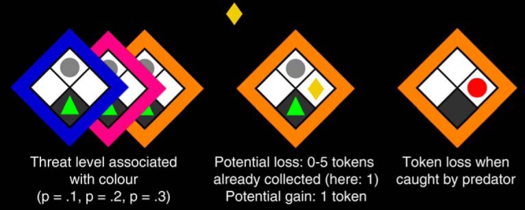
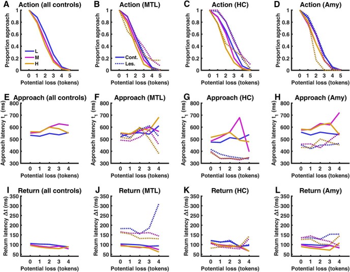
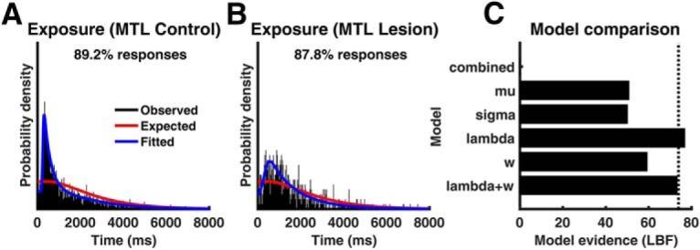
Anxiety comprises a suite of behaviors to deal with potential threat and is often modeled in approach-avoidance conflict tasks. Collectively, these tests constitute a predominant preclinical model of anxiety disorder. A body of evidence suggests that both ventral hippocampus and amygdala lesions impair anxiety-like behavior, but the relative contribution of these two structures is unclear. A possible reason is that approach-avoidance conflict tasks involve a series of decisions and actions, which may be controlled by distinct neural mechanisms that are difficult to disentangle from behavioral readouts. Here, we capitalize on a human approach-avoidance conflict test, implemented as computer game, that separately measures several action components. We investigate three patients of both sexes with unspecific unilateral medial temporal lobe (MTL) damage, one male with selective bilateral hippocampal (HC), and one female with selective bilateral amygdala lesions, and compare them to matched controls. MTL and selective HC lesions, but not selective amygdala lesions, increased approach decision when possible loss was high. In contrast, MTL and selective amygdala lesions, but not selective HC lesions, increased return latency. Additionally, selective HC and selective amygdala lesions reduced approach latency. In a task targeted at revealing subjective assumptions about the structure of the computer game, MTL and selective HC lesions impacted on reaction time generation but not on the subjective task structure. We conclude that deciding to approach reward under threat relies on hippocampus but not amygdala, whereas vigor of returning to safety depends on amygdala but not on hippocampus.SIGNIFICANCE STATEMENT Approach-avoidance conflict tests are widely investigated in rodents, and increasingly in humans, to understand the neural basis of anxiety-like behavior. However, the contribution of the most relevant brain regions, ventral hippocampus and amygdala, is incompletely understood. We use a human computerized test that separates different action components and find that hippocampus, but not amygdala, lesions impair approach decisions, whereas amygdala, but not hippocampus, lesions impair the vigor of return to safety.

Keywords: anxiety-like behavior; approach decision; approach–avoidance conflict; clinical lesion models; double-dissociation; escape vigor.


Figures

Figure 1.
Figure 1.
Behavioral task. On each trial, a human player (green triangle) rests in a safe place on the bottom of grid, while a “predator” is sleeping at the top (gray circle). On each epoch, up to six successive reward tokens (yellow rhombi) appear. To obtain a token, the player uses the left/right cursor keys to move out of the safe place and back. The colored frame indicates the threat level of the sleeping predator with color/threat association balanced across subjects. When caught, all tokens are lost. Potential loss is the number of tokens already collected on this epoch. (p: probability to get caught per 100 ms outside of safe place).
Figure 2.
Figure 2.
Behavioral results, displayed across the entire control group (A, E, I) and for each lesion type separately together with their respective control participants (B, F, J: MTL lesion; C, G, K: HC lesion; D, H, L: Amygdala lesion). Blue, Low threat level (L); purple, medium threat level (M); orange, high threat level (H). Solid lines, Control participants; dashed lines, patients. MTL, Surgical medial temporal lobe lesion; HC, selective bilateral hippocampus lesion; Amy, selective bilateral amygdala lesion. Approach and return are not displayed for the sixth token (potential loss 5 tokens) as this was rarely collected and therefore not included into statistical analysis.
Figure 3.
Figure 3.
Explicit memory of catch rate after the experiment. True catch rate depends on behavior and may change over the course of the experiment. For lesion patients, color/threat association was randomized, and each control participant was presented with the same association as their respective patient. Blue, Low threat level; purple, medium threat level; orange, high threat level. A: all control participants. B-D: lesion patients.
Figure 4.
Figure 4.
A, Fitted linear coefficients for the relationship between potential loss and proportion of approach, for the control and MTL lesion group (mean ± pooled SEM). BD, Fitted linear coefficients (B), mean approach latency (C), and mean return latency (D), for individual MTL patients (M1–M3), selective bilateral hippocampus lesion patient (HC), and selective bilateral amygdala lesion patient (Amy). Red dots indicate patients. *p < 0.05, one-tailed.
Figure 5.
Figure 5.
Exposure times from the safe-predator-exposure task for MTL lesion controls (A) and MTL lesion patients (B), with respect to the most recently appearing token. %Responses, Percentage of responses included in the plot; remaining responses were made before the first token occurred. Red lines show the expected exposure times under a uniform null distribution across the trial. C, Evidence for different models to distinguish controls and MTL patients expressed as LBF (larger is better) with respect to a reference model with no difference between the group (combined). Dashed line indicates decisiveness threshold (LBF difference >3).

References

    1. Amemori K, Amemori S, Graybiel AM (2015) Motivation and affective judgments differentially recruit neurons in the primate dorsolateral prefrontal and anterior cingulate cortex. J Neurosci 35:1939–1953. 10.1523/JNEUROSCI.1731-14.2015 - DOI - PMC - PubMed
    1. Bach DR. (2015) Anxiety-like behavioural inhibition is normative under environmental threat-reward correlations. PLoS Comput Biol 11:e1004646. 10.1371/journal.pcbi.1004646 - DOI - PMC - PubMed
    1. Bach DR. (2017) The cognitive architecture of anxiety-like behavioral inhibition. J Exp Psychol Hum Percept Perform 43:18–29. 10.1037/xhp0000282 - DOI - PMC - PubMed
    1. Bach DR, Talmi D, Hurlemann R, Patin A, Dolan RJ (2011) Automatic relevance detection in the absence of a functional amygdala. Neuropsychologia 49:1302–1305. 10.1016/j.neuropsychologia.2011.02.032 - DOI - PMC - PubMed
    1. Bach DR, Hurlemann R, Dolan RJ (2013) Unimpaired discrimination of fearful prosody after amygdala lesion. Neuropsychologia 51:2070–2074. 10.1016/j.neuropsychologia.2013.07.005 - DOI - PMC - PubMed

Publication types