. 2019 Sep 24;116(39):19695-19704.
doi: 10.1073/pnas.1903461116. Epub 2019 Sep 9.

Distinct intraspecies virulence mechanisms regulated by a conserved transcription factor

Affiliations

Distinct intraspecies virulence mechanisms regulated by a conserved transcription factor

James P R Connolly et al. Proc Natl Acad Sci U S A. .

Abstract

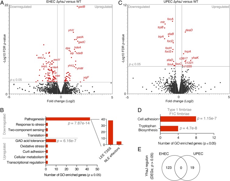
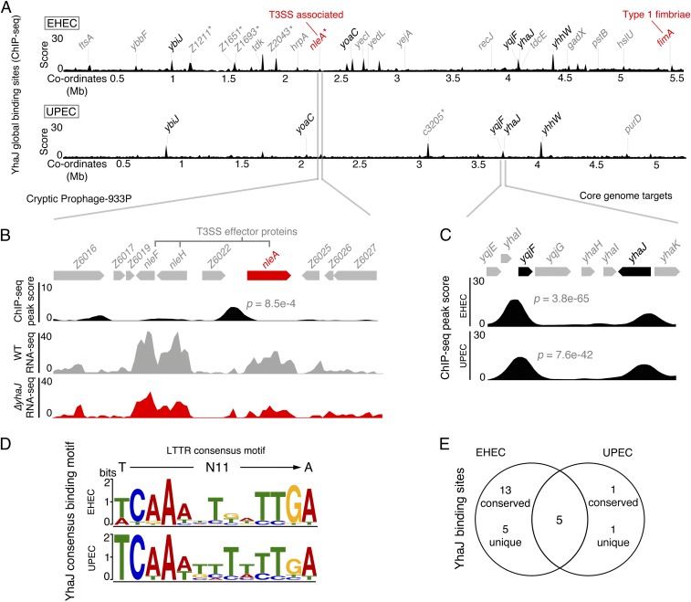
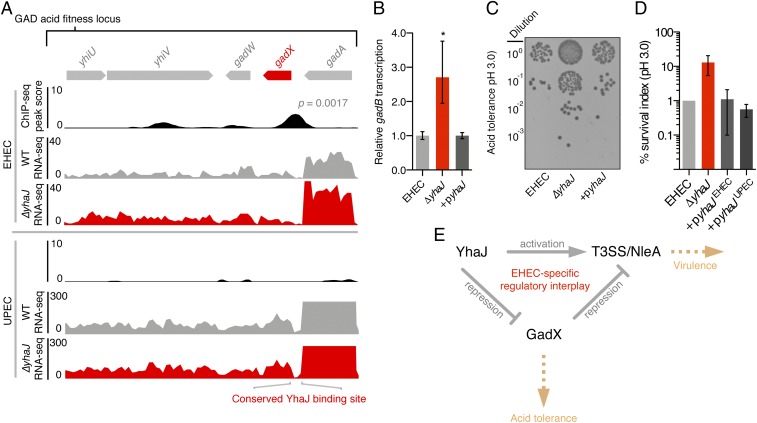
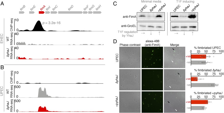
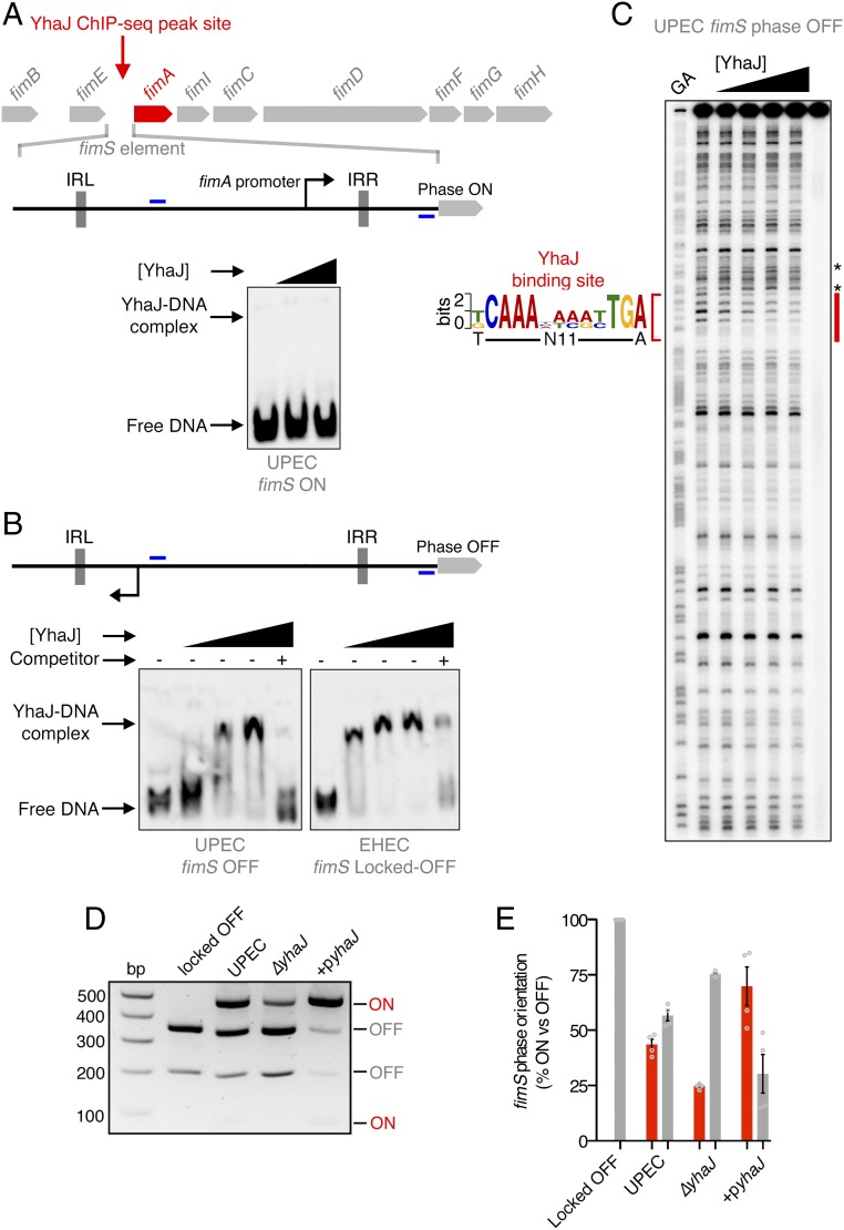
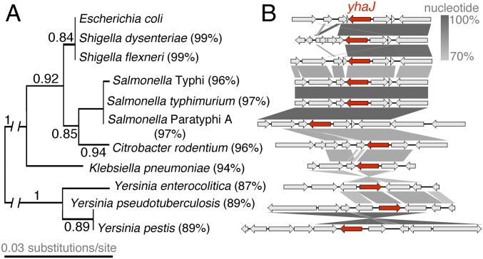
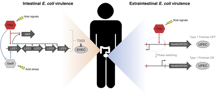
Tailoring transcriptional regulation to coordinate the expression of virulence factors in tandem with the core genome is a hallmark of bacterial pathogen evolution. Bacteria encode hundreds of transcription factors forming the base-level control of gene regulation. Moreover, highly homologous regulators are assumed to control conserved genes between members within a species that harbor the same genetic targets. We have explored this concept in 2 Escherichia coli pathotypes that employ distinct virulence mechanisms that facilitate specification of a different niche within the host. Strikingly, we found that the transcription factor YhaJ actively regulated unique gene sets between intestinal enterohemorrhagic E. coli (EHEC) and extraintestinal uropathogenic E. coli (UPEC), despite being very highly conserved. In EHEC, YhaJ directly activates expression of type 3 secretion system components and effectors. Alternatively, YhaJ enhances UPEC virulence regulation by binding directly to the phase-variable type 1 fimbria promoter, driving its expression. Additionally, YhaJ was found to override the universal GAD acid tolerance system but exclusively in EHEC, thereby indirectly enhancing type 3 secretion pleiotropically. These results have revealed that within a species, conserved regulators are actively repurposed in a "personalized" manner to benefit particular lifestyles and drive virulence via multiple distinct mechanisms.

Keywords: gene expression; niche; regulation; type 1 fimbriae; type 3 secretion.


Conflict of interest statement

The authors declare no conflict of interest.

Figures

Fig. 1.
Fig. 1.
YhaJ regulon is unique in 2 distinct E. coli pathotypes. (A) Volcano plot of RNA-seq transcriptome data displaying the pattern of gene expression values for ΔyhaJ relative to WT EHEC cultured in MEM-Hepes (data analyzed from ref. 20). Significantly differentially expressed genes (FDR-corrected P ≤ 0.05) are highlighted in red, with the gray lines representing the boundary for identification of up- or down-regulated genes. Selected genes related to the T3SS, acid tolerance, and known YhaJ targets are indicated. (B) Gene ontology analysis of EHEC DEGs identifying significantly enriched functional categories (P ≤ 0.05). The major groups are highlighted, and the Inset graph depicts the number of genes related to the LEE island or non–LEE-encoded effectors. (C) Volcano plot of RNA-seq transcriptome data displaying the pattern of gene expression values for ΔyhaJ versus WT UPEC cultured identically as EHEC. (D) Gene ontology analysis of UPEC DEGs with the class of adhesion genes highlighted above. (E) Venn diagram comparing the YhaJ regulon of EHEC and UPEC illustrating the lack of any commonality in significant DEGs. All transcriptome experiments were performed in biological triplicate.
Fig. 2.
Fig. 2.
YhaJ binds to unique core and horizontal chromosomal locations in distinct E. coli pathotypes. (A) Global chromosome binding dynamics of YhaJ in EHEC and UPEC identified by ChIP-seq. Significant peaks (P ≤ 0.01; 2 biological replicates) related to known targets are labeled in black, novel targets are in gray, and pathotype-specific targets are marked with an asterisk. The virulence-associated targets nleA (T3SS-associated) and fimA (type 1 fimbriae) are highlighted in red. (B) Expanded view of the horizontally acquired CP-933P locus in EHEC. The prophage region encodes multiple non–LEE-encoded effectors as indicated. The ChIP-seq track illustrates the identified YhaJ binding site upstream of nleA and RNA-seq data illustrate the downshift in transcription from nleA for ΔyhaJ (red) versus WT EHEC (gray). (C) Expanded view of ChIP-seq peaks identified for 2 common core genome targets, yhaJ itself and yqjF. The P values for yqjF peaks are indicated. (D) The computed consensus binding motif for YhaJ targets in EHEC and UPEC based on ChIP-seq data. The motifs match the generalized consensus sequence for LTTRs (T-N11-A) as illustrated above. (E) Comparison of YhaJ binding sites in EHEC and UPEC. ChIP-seq experiments were performed in biological duplicate.
Fig. 3.
Fig. 3.
YhaJ overrides the regulation of EHEC acid tolerance to control virulence via direct and indirect routes. (A) Expanded view of the EHEC YhaJ binding site upstream of the GAD acid tolerance regulator gene gadX. (A, Upper) RNA-seq tracks show the upshift in GAD gene transcription for ΔyhaJ (red) versus WT EHEC (gray). (A, Lower) Tracks reveal the lack of this regulation in a UPEC genetic background. The conserved sequence of the YhaJ-binding motif in both strains is illustrated (Bottom). (B) Repression of acid tolerance by YhaJ confirmed by qRT-PCR analysis of the GadX-regulated gene gadB in WT EHEC, ΔyhaJ, and +pyhaJ backgrounds. *P ≤ 0.05 derived from 3 biological replicates ±SD (Student’s t test). (C) Acid tolerance assay of WT EHEC, ΔyhaJ, and complemented (+pyhaJ) cells exposed to acidic conditions (pH 3.0). (D) Quantification of CFUs from B representing the mean survival relative to the WT (±SD of 4 biological replicates). The EHEC ΔyhaJ phenotype was complemented with both the EHEC and UPEC yhaJ alleles expressed in trans (+pyhaJEHEC and +pyhaJUPEC, respectively). (E) The regulatory interplay between YhaJ and GadX in fine-tuning T3SS expression in EHEC. Pointed and blunt arrows represent activation and repression, respectively, whereas the yellow arrows indicate the associated phenotype of each pathway.
Fig. 4.
Fig. 4.
YhaJ enhances expression of type 1 fimbriae in UPEC. (A) Expanded view of the YhaJ binding site upstream of the silent T1F locus in EHEC identified by ChIP-seq. The associated RNA-seq tracks highlight the lack of transcription from this silent locus in both WT EHEC (gray) and ΔyhaJ (red). (B) ChIP-seq data from the active T1F locus in UPEC illustrating the lack of YhaJ enrichment but a downshift in fimA transcription for ΔyhaJ (red) versus WT UPEC (gray). (C) Immunoblot analysis of FimA expression levels from WT UPEC, ΔyhaJ, and the complemented mutant +pyhaJ grown in minimal media (ChIP/RNA-seq conditions) and T1F-inducing conditions (static LB at 37 °C). Levels of GroEL were used to assess equal loading, and the influence of yhaJ mutation/complementation on T1F expression is highlighted (Bottom). Multiple biological replicates of immunoblots were performed. (D) Phase-contrast microscopy of WT UPEC, ΔyhaJ, and +pyhaJ grown under T1F-inducing conditions overlaid with immunofluorescence images of the cells probed using anti-FimA and Alexa-488 antibodies. The level of fimbriation around single cells was assessed (black arrows, T1F-positive; white arrows, T1F-negative) from more than 5 random fields of view per replicate and expressed as percentage fimbriated cells within the population (Right). Error bars represent SD, and the experiments were performed on 3 independent occasions.
Fig. 5.
Fig. 5.
YhaJ promotes activation of the fimS element by binding exclusively in the OFF orientation. (A) Schematic depiction of the phase-variable fimS element, containing an invertible promoter, in the ON orientation. EMSA analysis showing that YhaJ does not bind UPEC fimS in the ON orientation. (B) Schematic depiction of fimS in the OFF orientation. EMSAs demonstrating YhaJ binding over increasing concentrations to both UPEC and EHEC exclusively in the OFF orientation (Bottom). Excess unlabeled probe was added as a competitor to demonstrate binding specificity. Positions of primers used to amplify EMSA probes are indicated in blue. (C) DNaseI footprinting analysis of YhaJ bound to fimS. The protected region is indicated in red with the corresponding YhaJ/LTTR sequence motif indicated. The asterisks denote canonical hypersensitive sites flanking the protected region. (D) T1F phase-switching assay used to demonstrate YhaJ-activated inversion of fimS. The fimS locus contains a single HinfI restriction site, and DNA from cultures of WT UPEC, ΔyhaJ, and +pyhaJ was digested with HinfI and analyzed on 2% agarose. The banding pattern indicates the abundance of the population expressing T1F ON (red) or OFF (gray). WT EHEC which harbors a locked-OFF fimS was used as a control. (E) Quantification of T1F phase-switching assays illustrates the relative percentage of the population expressing fimS ON or OFF. Data represent the mean of 4 biological replicates ±SD.
Fig. 6.
Fig. 6.
YhaJ is highly conserved among prototypical gram-negative pathogens. (A) Maximum-likelihood tree generated from YhaJ-coding sequences of the prototypical gram-negative pathogens with the associated branch bootstrap fractions indicated. The percentage identity to the EHEC sequence as determined by BLASTp is indicated to the right of each species name. (B) Genomic context of yhaJ from the aforementioned isolates. The yhaJ ORF is indicated in red and all flanking genes are in gray. The gradient between schematics represents the percentage sequence similarity as indicated on the scale bar. Genes with no connecting gradient bar are unique to the context of each isolate.
Fig. 7.
Fig. 7.
Model of pleiotropic virulence regulation by YhaJ in distinct pathotypes. EHEC (intestinal niche) virulence is controlled by YhaJ pleiotropically—direct activation of the T3SS and prophage-encoded NleA, and indirect enhancement of T3SS by direct repression of GadX. UPEC (extraintestinal niche) virulence is enhanced by YhaJ through directly promoting phase ON orientation of the Fim switch element, leading to T1F expression.

References

    1. Browning D. F., Busby S. J. W., Local and global regulation of transcription initiation in bacteria. Nat. Rev. Microbiol. 14, 638–650 (2016). - PubMed
    1. Balleza E., et al. , Regulation by transcription factors in bacteria: Beyond description. FEMS Microbiol. Rev. 33, 133–151 (2009). - PMC - PubMed
    1. Seshasayee A. S. N., Sivaraman K., Luscombe N. M., “An overview of prokaryotic transcription factors” in A Handbook of Transcription Factors, Hughes T., Ed. (Springer, 2011), vol. 52, pp. 7–23. - PubMed
    1. Perez J. C., Groisman E. A., Evolution of transcriptional regulatory circuits in bacteria. Cell 138, 233–244 (2009). - PMC - PubMed
    1. Moran N. A., Microbial minimalism: Genome reduction in bacterial pathogens. Cell 108, 583–586 (2002). - PubMed

Publication types

MeSH terms

LinkOut - more resources