. 2019 Sep 9;10(9):646.
doi: 10.1038/s41419-019-1887-4.

Direct neuronal reprogramming of olfactory ensheathing cells for CNS repair

Affiliations

Direct neuronal reprogramming of olfactory ensheathing cells for CNS repair

Xiu Sun et al. Cell Death Dis. .

Abstract

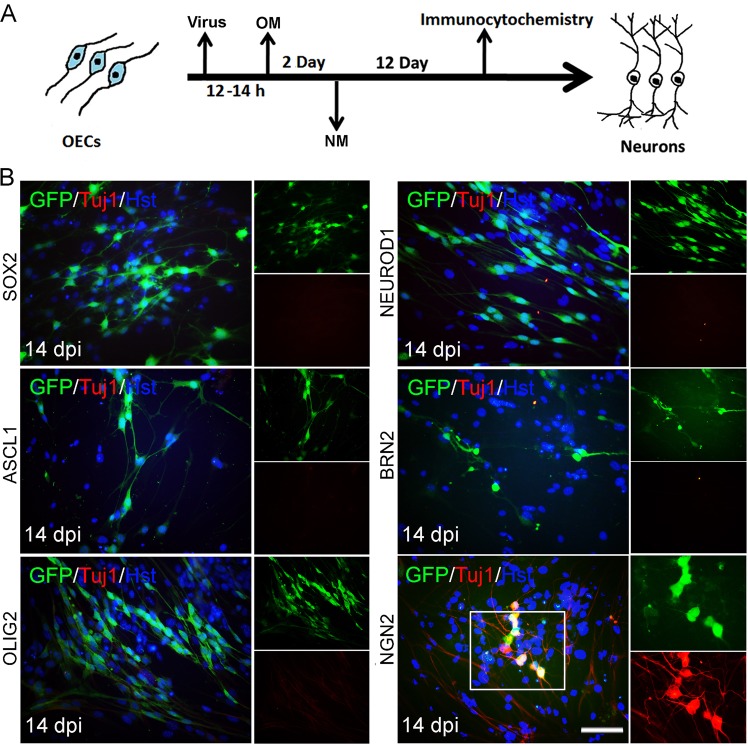
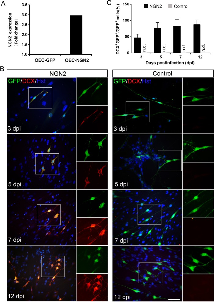
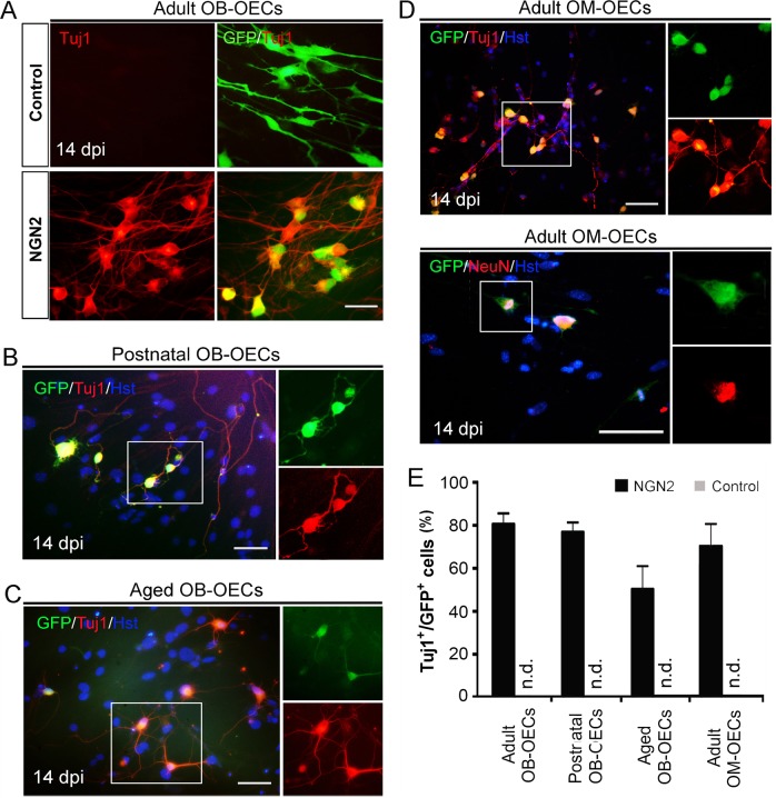
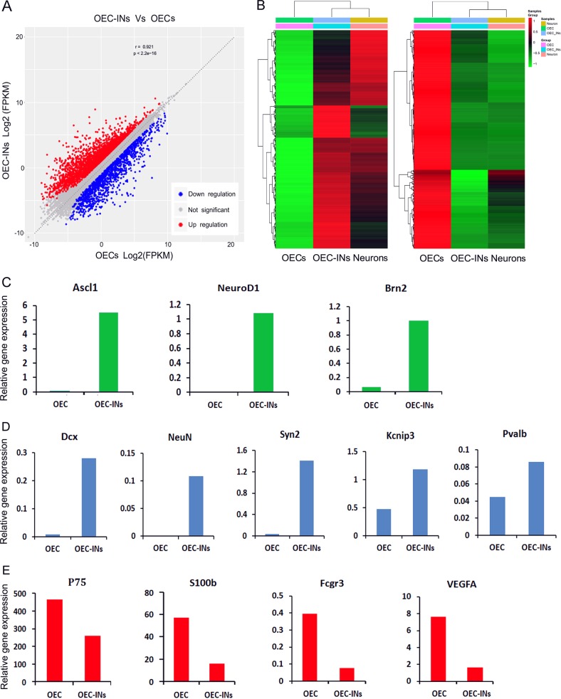
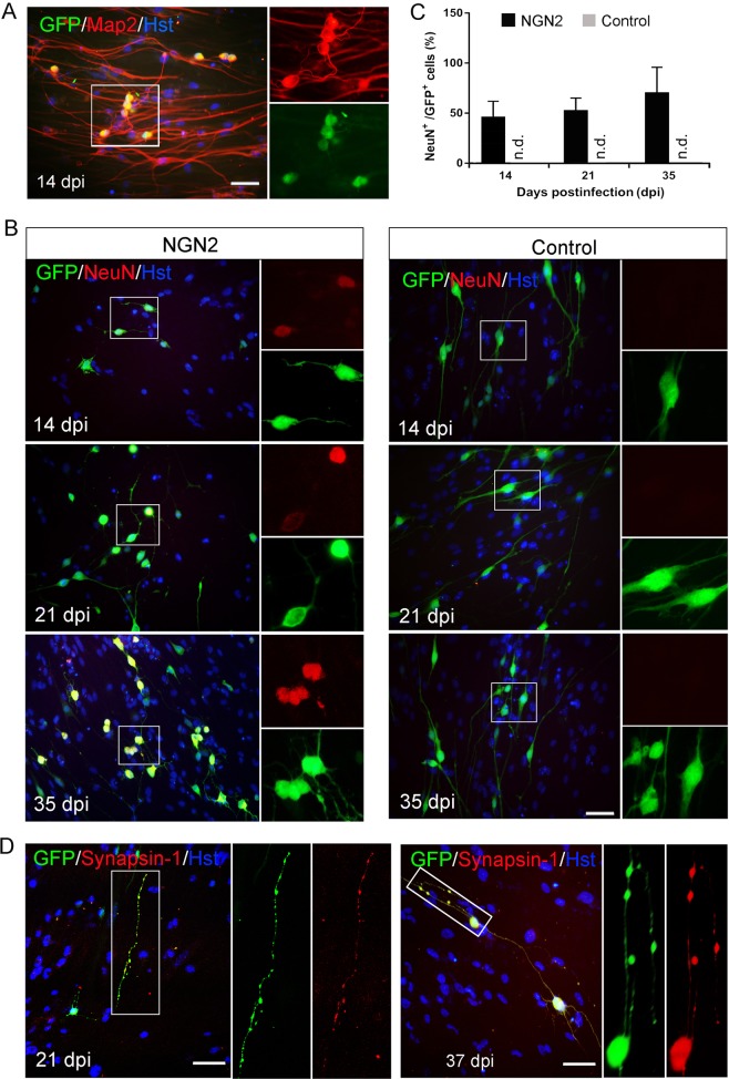
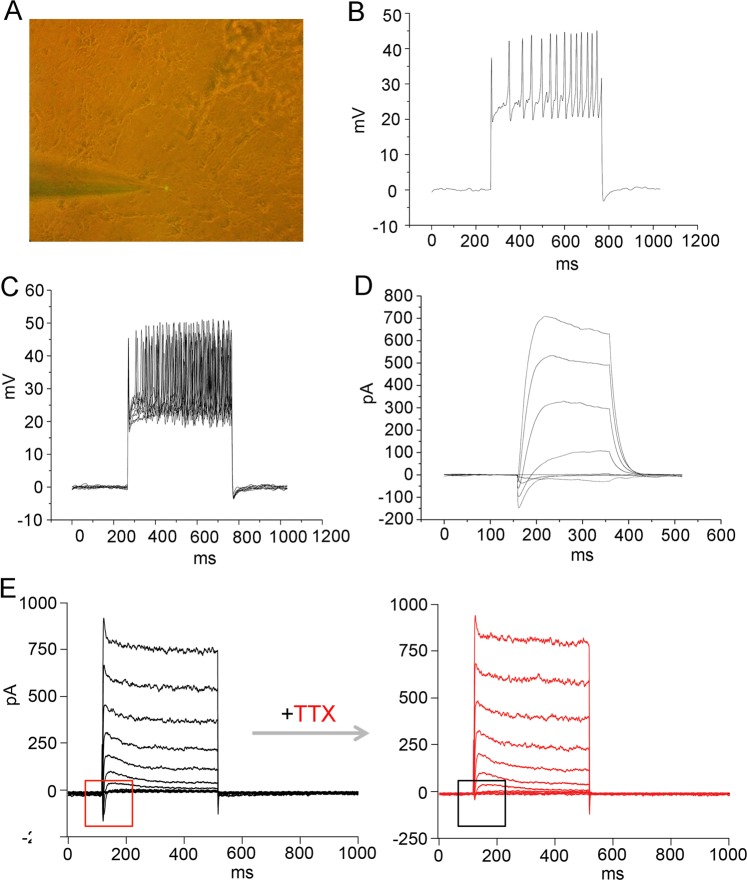
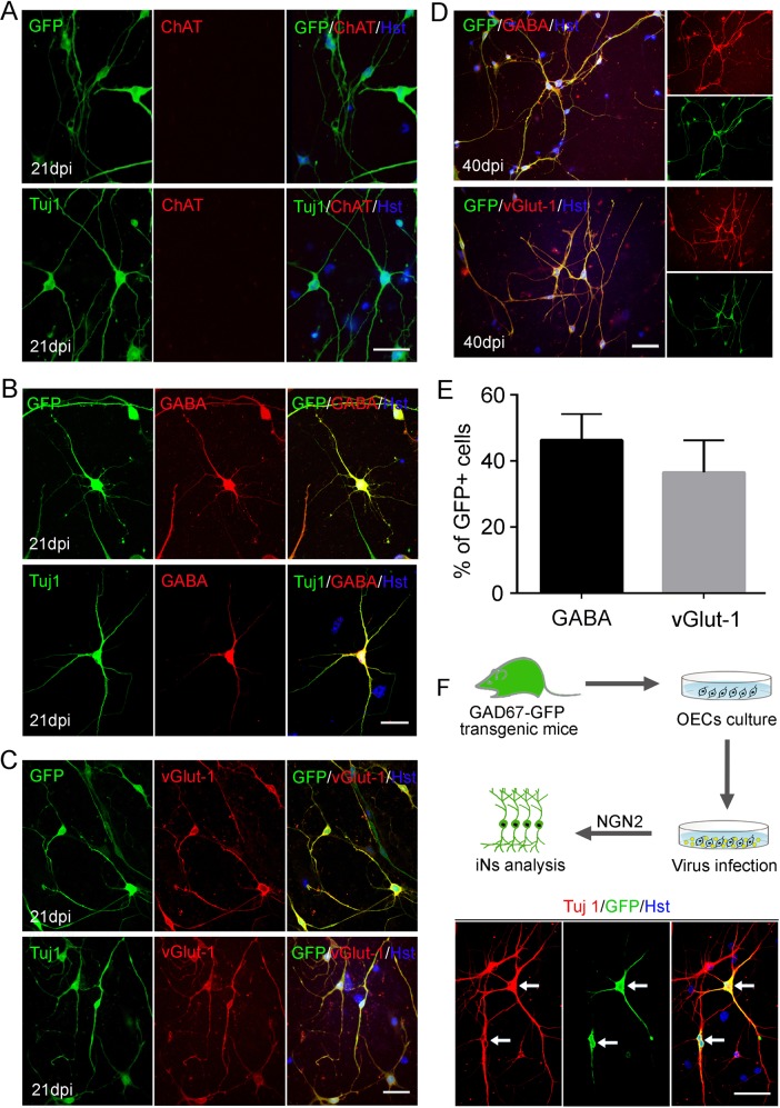
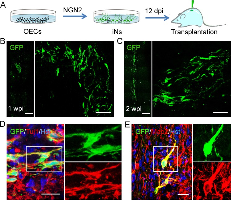
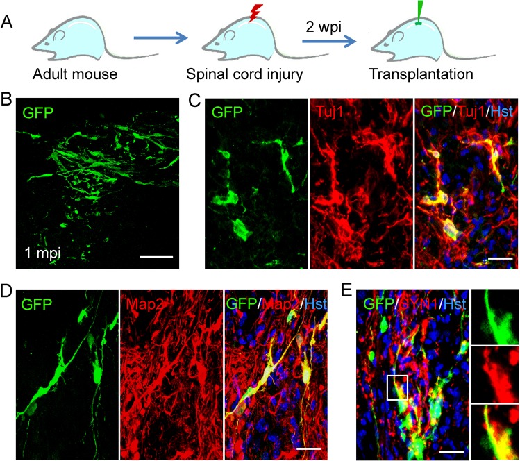
Direct conversion of readily available non-neural cells from patients into induced neurons holds great promise for neurological disease modeling and cell-based therapy. Olfactory ensheathing cells (OECs) is a unique population of glia in olfactory nervous system. Based on the regeneration-promoting properties and the relative clinical accessibility, OECs are attracting increasing attention from neuroscientists as potential therapeutic agents for use in neural repair. Here, we report that OECs can be directly, rapidly and efficiently reprogrammed into neuronal cells by the single transcription factor Neurogenin 2 (NGN2). These induced cells exhibit typical neuronal morphologies, express multiple neuron-specific markers, produce action potentials, and form functional synapses. Genome-wide RNA-sequencing analysis shows that the transcriptome profile of OECs is effectively reprogrammed towards that of neuronal lineage. Importantly, these OEC-derived induced neurons survive and mature after transplantation into adult mouse spinal cords. Taken together, our study provides a direct and efficient strategy to quickly obtain neuronal cells from adult OECs, suggestive of promising potential for personalized disease modeling and cell replacement-mediated therapeutic approaches to neurological disorders.


Conflict of interest statement

The authors declare that they have no conflict of interest.

Figures

Fig. 1
Fig. 1. Induction of neuronal fate on OECs.
a Experimental scheme of transcription factor-mediated neuronal reprogramming from OECs. OM, OEC culture medium; NM, neuronal induction medium. b Immunocytochemical analysis of induced neurons from OECs by ectopic expression of individual transcription factors, using antibody against Tuj1. Scale bar = 50 μm
Fig. 2
Fig. 2. NGN2-induced lineage reprogramming of OECs into DCX+ cells.
a RT-PCR confirming NGN2 expression in the virus-transduced OECs. b Representative micrographs of NGN2-induced neurons from OECs by staining with DCX at different time points. c Quantification of induced DCX+ cells during the indicated time course (n = 20 random fields from triplicate samples; n.d., not detected). Scale bar = 50 μm
Fig. 3
Fig. 3. NGN2-induced neuronal reprogramming from OECs of different origins.
a–d Immunocytochemical analysis of lineage reprogramming of OECs from adult olfactory bulb (Adult OB-OECs) (a), postnatal olfactory bulb (Postnatal OB-OECs) (b), aged olfactory bulb (Aged OB-OECs) (c), and adult olfactory mucosa (Adult OM-OECs) (d) by staining with antibodies against Tuj1 and NeuN at 14 dpi. e NGN2-induced neuronal reprogramming efficiency of OECs of different origins (n = 20 random fields from triplicate samples; n.d., not detected). Scale bar = 50 μm
Fig. 4
Fig. 4. Transcriptome analysis of induced neuronal cells from OECs.
a Scatterplots comparing gene expression levels between OEC-derived induced neurons (OEC-INs) and OECs (OECs). Up-regulation genes were highlighted in red; down regulation genes were highlighted in blue. b Heatmap of genes differentially expressed in RNA-sequencing analysis performed on OECs, OEC-INs, and cortical neurons (Neurons). Compared with OECs, the up-regulation genes (left panel) and down regulation genes (right panel) were identified in OEC-INs. ce Gene expression (FPKM) of representative genes for neurogenic factors (c), neuronal lineage markers (d), and OEC markers (e), in OECs and OEC-INs at 14 dpi
Fig. 5
Fig. 5. Immunocytochemical analysis of NGN2-mediated neuronal conversion process.
a Lack of marker expression for neural progenitors during the reprogramming process. The expression in neural stem cells (NSCs) served as a positive control. b Time-course analysis of proliferating cells by staining with PCNA (n = 20 randomly selected fields from triplicate samples). c, d Incorporation of BrdU in NGN2-induced neuronal cells. Cells were treated with BrdU for the indicated duration after viral infection (b). OM, OEC culture medium; NM, neuronal induction medium. *P < 0.05, ***P < 0.001 by Student’s t-tests. Scale bar = 50 μm
Fig. 6
Fig. 6. Maturation of NGN2-induced neurons from OECs.
a Representative micrographs of mature NGN2-induced neurons from adult OECs by staining with mature neuronal marker Map2 at 14 dpi. b, c Time-course analysis of the maturation of induced neuronal cells by staining with mature neuronal marker NeuN (n = 20 randomly selected fields from triplicate samples). d Expression of the presynaptic marker synapsin-1 in the converted cells at 21 or 37 dpi. Scale bar = 50 μm
Fig. 7
Fig. 7. Electrophysiological properties of NGN2-induced neurons from OECs.
a A representative visual field of the patched neuron containing GFP fluorescence. b Single action potential (AP) firing of an induced neuron which was co-cultured with astrocytes for 34 days in response to injection of a suprathreshold current pulse. c Multiple AP firing elicited by repetitious depolarizing current stimuli. d Voltage-clamp traces showing fast inward current and persistent outward current on depolarization. e Tetrodotoxin (TTX)-sensitive sodium currents. The voltage-gated Na+ currents recorded from an induced neuron (left panel) and subsequently blocked by TTX (right panel)
Fig. 8
Fig. 8. Subtype analysis of NGN2-induced neurons from OECs.
a No expression of ChAT was detected in the induced neurons at 21 dpi. b, c Ectopic NGN2-induced OECs reprogramming into GABA+ (b) or vGlut1+ (c) neurons at 21 dpi. d Representative images of NGN2-induced GABA+ or vGlut1+ neurons at 40 dpi. e Quantification of the percentage of GFP+ cells expressing GABA or vGlut1 over the total of GFP+ cells (n = 20 randomly selected fields from triplicate samples). f Induction of GABAergic neurons from OECs of GAD67-GFP transgenic mice. OECs were cultured from olfactory bulb of adult GAD67-GFP transgenic mice, and then infected with NGN2-expressing lentivirus (without co-expressing GFP) to induce neuronal reprogramming. Immunocytochemical analysis was performed at 21 dpi. Scale bar = 50 μm
Fig. 9
Fig. 9. Transplantation of OEC-converted neurons into normal adult spinal cord.
a Schematic diagram showing the experimental procedure of intraspinal injection of induced cells. b, c Survival of GFP-labeled induced cells at 1 and 2 weeks post injection (wpi). d, e Immunohistochemical analysis of OEC-derived neurons by staining with antibodies against Tuj1 and Map2 in the spinal cord of NOD-SCID mice. Scale bar, 100 μm for b, c; 50 μm for d, e
Fig. 10
Fig. 10. Transplantation of OEC-converted neurons into injured adult spinal cord.
a Schematic diagram showing the experimental procedure of stereotactical injection of induced cells into crushed spinal cord. b Survival of GFP-labeled induced cells at one month post injection (mpi). ce Immunohistochemical analysis of OEC-derived neurons by staining with antibodies against Tuj1, Map2, and synapsin-1 (SYN1) in the spinal cord of NOD-SCID mice. Scale bar, 100 μm for b; 50 μm for ce

References

    1. Dong Z, Pan K, Pan J, Peng Q, Wang Y. The possibility and molecular mechanisms of cell pyroptosis after cerebral ischemia. Neurosci. Bull. 2018;34:1131–1136. doi: 10.1007/s12264-018-0294-7. - DOI - PMC - PubMed
    1. Case LC, Tessier-Lavigne M. Regeneration of the adult central nervous system. Curr. Biol. 2005;15:R749–R753. doi: 10.1016/j.cub.2005.09.008. - DOI - PubMed
    1. Fitch MT, Silver J. CNS injury, glial scars, and inflammation: Inhibitory extracellular matrices and regeneration failure. Exp. Neurol. 2008;209:294–301. doi: 10.1016/j.expneurol.2007.05.014. - DOI - PMC - PubMed
    1. Takahashi K, Yamanaka S. Induction of pluripotent stem cells from mouse embryonic and adult fibroblast cultures by defined factors. Cell. 2006;126:663–676. doi: 10.1016/j.cell.2006.07.024. - DOI - PubMed
    1. Marro S, et al. Direct lineage conversion of terminally differentiated hepatocytes to functional neurons. Cell Stem Cell. 2011;9:374–382. doi: 10.1016/j.stem.2011.09.002. - DOI - PMC - PubMed

Publication types