

IFN-γ restores the impaired function of RNase L and induces mitochondria-mediated apoptosis in lung cancer
- PMID: 31501431
- PMCID: PMC6733796
- DOI: 10.1038/s41419-019-1902-9
IFN-γ restores the impaired function of RNase L and induces mitochondria-mediated apoptosis in lung cancer
Abstract
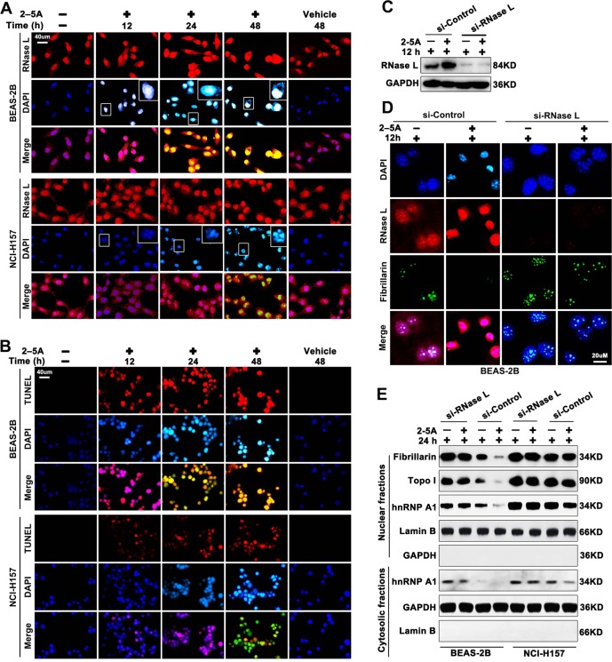
RNase L is an essential component in interferon (IFN)-mediated antiviral signaling that showed antitumor effects in cancer. Cancer immunotherapy based on interferon has achieved encouraging results that indicate an applicable potential for cancer therapy. Here we showed that function of RNase L, though highly upregulated, was functionally impaired both in nuclear and cytoplasm in lung cancer cells. In normal lung epithelial cells, RNase L activation induced by 2-5A promoted nuclear condensation, DNA cleavage, and cell apoptosis, while in lung cancer cells, these processes were inhibited and RNase L-mediated downregulation of fibrillarin, Topo I and hnRNP A1 was also impaired in lung cancer cells. Moreover, the impairment of RNase L in lung cancer cells was due to the elevated expression of RLI. Application of IFN-γ to lung cancer cells led to enhanced expression of RNase L that compensated the RLI inhibition and restored the cytoplasmic and nuclear function of RNase L, leading to apoptosis of lung cancer cells. Thus, the present study discovered the impaired function and mechanism of RNase L in lung cancer cells and proved the efficacy of IFN-γ in restoring RNase L function and inducing apoptosis in the lung cancer cell. These results indicated the RNase L as a therapeutic target in lung cancer cells and immunotherapy of IFN-γ may serve as an adjuvant to enhance the efficacy.
Conflict of interest statement
The authors declare that they have no conflict of interest.
Figures


Similar articles
-
Induction of indoleamine 2,3-dioxygenase (IDO) enzymatic activity contributes to interferon-gamma induced apoptosis and death receptor 5 expression in human non-small cell lung cancer cells.Asian Pac J Cancer Prev. 2014;15(18):7995-8001. doi: 10.7314/apjcp.2014.15.18.7995. Asian Pac J Cancer Prev. 2014. PMID: 25292102
-
RNase L Amplifies Interferon Signaling by Inducing Protein Kinase R-Mediated Antiviral Stress Granules.J Virol. 2020 Jun 16;94(13):e00205-20. doi: 10.1128/JVI.00205-20. Print 2020 Jun 16. J Virol. 2020. PMID: 32295917 Free PMC article.
-
The association of elevated 2',5'-oligoadenylate-dependent RNase L with lung cancer correlated with deficient enzymatic activity and decreased capacity of RNase L dimerization.Lung Cancer. 2012 Oct;78(1):30-8. doi: 10.1016/j.lungcan.2012.07.010. Epub 2012 Aug 24. Lung Cancer. 2012. PMID: 22925698
-
RNase L inhibitor (RLI) antisense constructions block partially the down regulation of the 2-5A/RNase L pathway in encephalomyocarditis-virus-(EMCV)-infected cells.Eur J Biochem. 1998 Jun 1;254(2):248-55. doi: 10.1046/j.1432-1327.1998.2540248.x. Eur J Biochem. 1998. PMID: 9660177
-
The 2-5A system in viral infection and apoptosis.Biomed Pharmacother. 1998;52(9):386-90. doi: 10.1016/s0753-3322(99)80006-7. Biomed Pharmacother. 1998. PMID: 9856285 Review.
Cited by
-
Fishing for newly synthesized proteins with phosphonate-handles.Nat Commun. 2020 Jun 26;11(1):3244. doi: 10.1038/s41467-020-17010-0. Nat Commun. 2020. PMID: 32591520 Free PMC article.
-
Eosinophil and IFN-γ associated with immune-related adverse events as prognostic markers in patients with non-small cell lung cancer treated with immunotherapy.Front Immunol. 2023 Mar 6;14:1112409. doi: 10.3389/fimmu.2023.1112409. eCollection 2023. Front Immunol. 2023. PMID: 36949952 Free PMC article.
-
MiR-302a Regenerates Human Corneal Endothelial Cells against IFN-γ-Induced Cell Death.Cells. 2022 Dec 22;12(1):36. doi: 10.3390/cells12010036. Cells. 2022. PMID: 36611829 Free PMC article.
-
5-Aminolevulinic acid/sodium ferrous citrate enhanced the antitumor effects of programmed cell death-ligand 1 blockade by regulation of exhausted T cell metabolism in a melanoma model.Cancer Sci. 2021 Jul;112(7):2652-2663. doi: 10.1111/cas.14930. Epub 2021 May 22. Cancer Sci. 2021. PMID: 33934440 Free PMC article.
-
Activation of the viral sensor oligoadenylate synthetase 2 (Oas2) prevents pregnancy-driven mammary cancer metastases.Breast Cancer Res. 2022 May 3;24(1):31. doi: 10.1186/s13058-022-01525-z. Breast Cancer Res. 2022. PMID: 35505346 Free PMC article.
References
-
- Dayal Shubham, Zhou Jun, Manivannan Praveen, Siddiqui Mohammad, Ahmad Omaima, Clark Matthew, Awadia Sahezeel, Garcia-Mata Rafael, Shemshedini Lirim, Malathi Krishnamurthy. RNase L Suppresses Androgen Receptor Signaling, Cell Migration and Matrix Metalloproteinase Activity in Prostate Cancer Cells. International Journal of Molecular Sciences. 2017;18(3):529. doi: 10.3390/ijms18030529. - DOI - PMC - PubMed
Publication types
MeSH terms
Substances
LinkOut - more resources
Full Text Sources
Medical
