. 2019 Sep 9;9(1):12910.
doi: 10.1038/s41598-019-49402-8.

A first experience of transduction for differentiated HepaRG cells using lentiviral technology

Affiliations

A first experience of transduction for differentiated HepaRG cells using lentiviral technology

Adeline Pivert et al. Sci Rep. .

Erratum in

Abstract

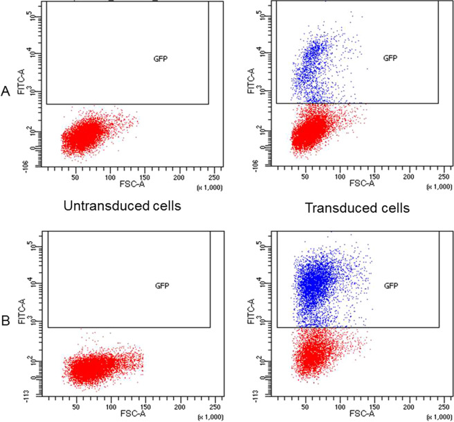
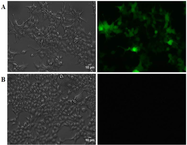
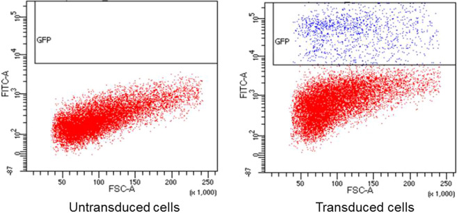
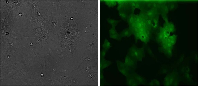
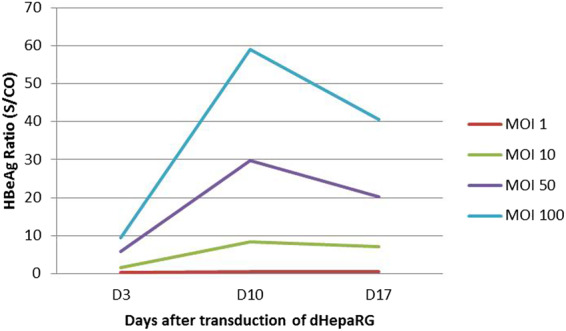
Currently, there is a lack of systems for studying the role of hepatitis B viral proteins, such as HBeAg and HBcAg, on liver injury. It is necessary to develop an original tool in order to clarify the role of these viral proteins in hepatic stellate cell activation, and to understand the molecular mechanisms of liver injury. HepaRG are the most reliable hepatocyte-like cells for studying liver functions or disorders. In this paper, we demonstrate that the transduction of differentiated HepaRG (dHepaRG) cells can be performed successfully using lentiviral particles. The production of a functional Green Fluorescent Protein (GFP) assessed by Fluorescence Activated Cell Sorting and fluorescence microscopy is up to 16% of GFP positive cells using a multiplicity of infection (MOI) of 2.4. We demonstrate that this technology can allow the stable expression of GFP during the long lifecycle of the cell (up to four weeks after the cell's passage). With this innovative tool, we aim to express viral proteins such as HBeAg or HBcAg in dHepaRG cells. The preliminary results of this work shows that HBeAg can be efficiently produced in dHepaRG cells and that increased MOI allows a better production of this protein. Our future objective will be to study the role of HBc and HBe proteins on the induction of hepatic fibrosis.


Conflict of interest statement

The authors declare no competing interests.

Figures

Figure 1
Figure 1
Quantification of GFP expression by flow cytometry in HEK 293T cells following lentiviral transduction in order to establish the functional titer of lentiviral particles. The horizontal axis represents the forward scatter size (FSC-A) where an increased signal may indicate an increase in cell size or budding. The vertical axis indicates the GFP fluorescence intensity (FITC-A). Blue dots represent GFP-positive cells. Red dots represent GFP-negative cells. The threshold for GFP positivity was determined according to negative control (non-transduced cells). The concentrated viral supernatant titer was calculated using various supernatant dilutions, an average GFP-positive cell count of 15.8%, corresponding to a 10−1 dilution of the viral supernatant (A). The percentage of GFP-positive cells using 10 μL of concentrated viral supernatant is 52.8% (B). Four assays were performed with the 10−1 dilution of the viral supernatant: the mean titer was 1.7 × 107 ± 1.05 × 107 TU/mL.
Figure 2
Figure 2
Phase contrast and fluorescence microscopy of 293T cells after transduction with lentiviral particles containing the GFP gene. Cells were transduced with 10 μL of concentrated viral supernatant and emitted a fluorescent signal (A); Non-transduced cells were used as negative control (B). The scale bar represents 10 μm (A,B).
Figure 3
Figure 3
Quantification of GFP expression by flow cytometry in dHepaRG cells 10 days after lentiviral transduction. The horizontal axis represents the forward scatter size, (FSC-A) where an increased signal may indicate an increase in cell size or budding. The vertical axis indicates the intensity of GFP fluorescence (FITC-A). Blue dots represent the cells expressing GFP. Red dots represent the cells that do not express GFP. The GFP positivity threshold of the cells was determined according to negative control (non-transduced cells). The percentage of GFP-positive cells at an MOI of 2.4 is 10%.
Figure 4
Figure 4
Phase contrast and fluorescence microscopy of dHepaRG cells expressing GFP 10 days after lentiviral transduction. Cells were transduced with viral supernatant at a MOI of 2.4. The scale bar represents 10 μm.
Figure 5
Figure 5
Phase contrast and fluorescence microscopy of dHepaRG cells expressing GFP 4 weeks after lentiviral transduction. Cells were transduced with viral supernatant at a MOI of 2.4. The scale bar represents 10 μm.
Figure 6
Figure 6
HBeAg ratio measured by CMIA for the different MOI tested at days 3, 10 and 17 after the transduction of dHepaRG (n = 1).

References

    1. Mormone E, George J, Nieto N. Molecular pathogenesis of hepatic fibrosis and current therapeutic approaches. Chem Biol Interact. 2011;193:225–231. doi: 10.1016/j.cbi.2011.07.001. - DOI - PMC - PubMed
    1. Liu W, et al. Hepatitis B virus core protein inhibits Fas-mediated apoptosis of hepatoma cells via regulation of mFas/FasL and sFas expression. FASEB J. 2014;29:1113–1123. doi: 10.1096/fj.14-263822. - DOI - PubMed
    1. Chen HY, Chen ZX, Huang RF, Lin N, Wang XZ. Hepatitis B virus X protein activates human hepatic stellate cells through upregulating TGFbeta1. Genet Mol Res. 2014;13:8645–8656. doi: 10.4238/2014.October.27.4. - DOI - PubMed
    1. Gripon P, et al. Infection of a human hepatoma cell line by hepatitis B virus. Proc Natl Acad Sci USA. 2002;99:15655–15660. doi: 10.1073/pnas.232137699. - DOI - PMC - PubMed
    1. Yan, H. et al. Sodium taurocholate cotransporting polypeptide is a functional receptor for human hepatitis B and D virus. Elife3 (2012). - PubMed