. 2019 Oct;16(10):987-990.
doi: 10.1038/s41592-019-0548-y. Epub 2019 Sep 9.

High-definition spatial transcriptomics for in situ tissue profiling

Affiliations

High-definition spatial transcriptomics for in situ tissue profiling

Sanja Vickovic et al. Nat Methods. 2019 Oct.

Abstract

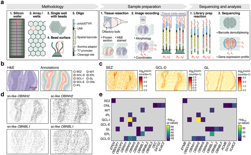
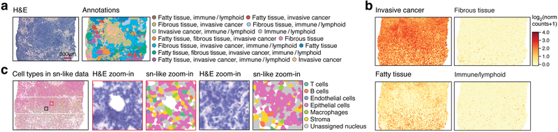
Spatial and molecular characteristics determine tissue function, yet high-resolution methods to capture both concurrently are lacking. Here, we developed high-definition spatial transcriptomics, which captures RNA from histological tissue sections on a dense, spatially barcoded bead array. Each experiment recovers several hundred thousand transcript-coupled spatial barcodes at 2-μm resolution, as demonstrated in mouse brain and primary breast cancer. This opens the way to high-resolution spatial analysis of cells and tissues.


Conflict of interest statement

Competing interests

F.S., J.F., J.L. and P.L.S. are authors on patents PCT/EP2012/056823 (WO2012/140224), PCT/EP2013/071645 (WO2014/060483) and PCT/EP2016/057355 applied for by Spatial Transcriptomics AB (10x Genomics) covering the described technology. M.R. is employed by Illumina Inc. A.R. is a founder and equity holder of Celsius Therapeutics and an SAB member of Syros Pharmaceuticals and Thermo Fisher Scientific.

Figures

Figure 1.
Figure 1.. High-definition spatial transcriptomics (HDST).
(a) HDST workflow. (b) Labeling of morphological layers. HDST H&E image of a main olfactory bulb and matching HDST (x,y) barcodes annotated into 9 morphological areas. (c) Layer-specific DE patterns in HDST. Shown is the summed normalized expression of positively enriched signature genes significantly (FDR<0.1, two-sided t-test) associated with each layer as annotated in (b). (d, e) Nuclei segmentation and binning of HDST as in (b). (d) Segmented nuclei (“sn-like”) and lightly binned (“sc-like”) spatial barcodes assigned (black) to each of two cell types as in (b). (e) Enrichment of sn- and sc-like spatial barcodes with assigned cell types (columns) to morphological layers (rows) as in (b). −log10(p-value) (one-sided Fisher’s exact test, Bonferroni adjusted, p-value<0.01) represents the color bar and grey tiles non-significant values.
Figure 2.
Figure 2.. HDST distinguishes cell types and niches in a breast cancer resection.
(a) Labeling of morphological layers. HDST H&E image (left) of a breast cancer section and matching HDST (x,y) barcodes annotated into 13 morphological areas (right, color code). (b) Layer-specific spatial DE patterns in HDST. Summed normalized expression of positively enriched signature genes significantly (FDR<0.1, two-sided t-test) associated with each layer as in (a). (c) Cell type assignments by single nuclei as in (a). Two zoomed in regions (black and red squares) with H&E and color coded segments.

References

    1. Oh SW et al. Nature 508, 207–214 (2014). - PMC - PubMed
    1. Lein E, Borm LE & Linnarsson S Science 358, 64–69 (2017). - PubMed
    1. Macosko EZ et al. Cell 161, 1202–1214 (2015). - PMC - PubMed
    1. Zheng GXY et al. Nat. Commun 8, 14049 (2017). - PMC - PubMed
    1. van den Brink SC et al. Nat. Methods 14, 935–936 (2017). - PubMed

Publication types