. 2019 Oct;37(10):1131-1136.
doi: 10.1038/s41587-019-0223-y. Epub 2019 Sep 9.

Supercooling extends preservation time of human livers

Affiliations

Supercooling extends preservation time of human livers

Reinier J de Vries et al. Nat Biotechnol. 2019 Oct.

Abstract

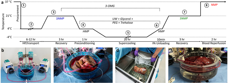
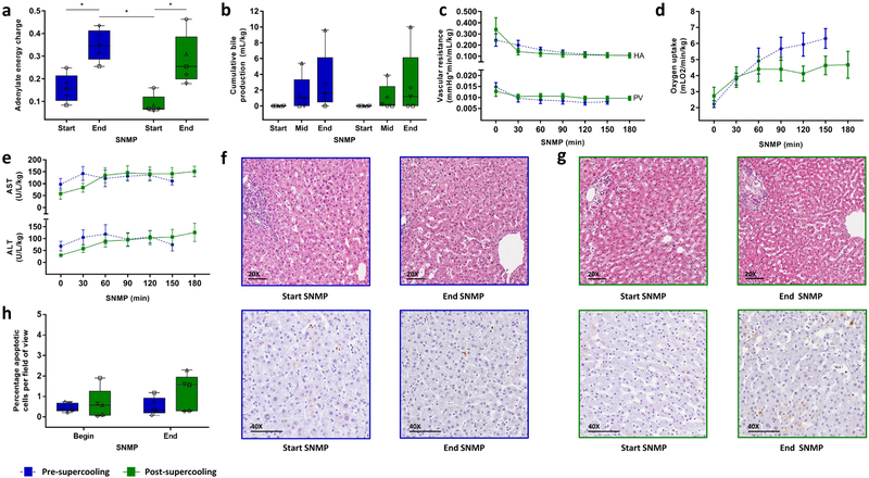
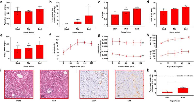
The inability to preserve vascular organs beyond several hours contributes to the scarcity of organs for transplantation1,2. Standard hypothermic preservation at +4 °C (refs. 1,3) limits liver preservation to less than 12 h. Our group previously showed that supercooled ice-free storage at -6 °C can extend viable preservation of rat livers4,5 However, scaling supercooling preservation to human organs is intrinsically limited because of volume-dependent stochastic ice formation. Here, we describe an improved supercooling protocol that averts freezing of human livers by minimizing favorable sites of ice nucleation and homogeneous preconditioning with protective agents during machine perfusion. We show that human livers can be stored at -4 °C with supercooling followed by subnormothermic machine perfusion, effectively extending the ex vivo life of the organ by 27 h. We show that viability of livers before and after supercooling is unchanged, and that after supercooling livers can withstand the stress of simulated transplantation by ex vivo normothermic reperfusion with blood.


Conflict of interest statement

The authors declare competing financial interests. Drs. Toner, Yarmush, de Vries, Uygun and Tessier have provisional patent applications relevant to this study. Dr. Uygun has a financial interest in Organ Solutions, a company focused on developing organ preservation technology. Author’s interests are managed by the MGH and Partners HealthCare in accordance with their conflict of interest policies.

Figures

Figure 1
Figure 1. Outline of research design
(a) Schematic temperature profile of the supercooling protocol: The overall research design, entails 8 steps. 1) Five human livers, rejected for transplantation, were procured in standard fashion and 2) transported under hypothermic preservation (HP) conditions. 3) Upon arrival, we recovered the grafts from the incurred warm and cold ischemia and collected pre-supercooling viability parameters during 3 hours of subnormothermic machine perfusion (SNMP). We supplemented the perfusate with 19.42 g/L (200 mM) 3-O-methyl-d-glucose (3-OMG) during the last hour of perfusion. 4) At the end of SNMP, we gradually lowered the perfusion temperature, which was followed by hypothermic machine perfusion (HMP) with University of Wisconsin solution (UW) supplemented with 50 g/L (1.43 μM) 35kD polyethylene glycol (PEG), 37.83 g/L (100 mM) trehalose dihydrate and 125.7 g/L (1.36 M) glycerol. 5) Following preconditioning with the protective agents, the livers were supercooled and stored free of ice at −4°C for 20 hours. 6) After supercooling, the protective agents were gradually washed out, and 7) the livers were recovered by SNMP, identical to pre-supercooling conditions except addition of Trolox to the perfusate and absence of 3-OMG and cooling at the end of SNMP. Post-supercooling viability parameters were collected during SNMP and compared to their baseline values. 8) Three livers were additionally reperfused with non-leukoreduced red blood cells and plasma at 37°C as a model for transplantation. (b) Machine perfusion system. (c) Liver during SNMP recovery. (d) Liver in supercooling basin of the chiller. (e) Normothermic reperfusion with blood of the supercooled liver.
Figure 2
Figure 2. Key ex vivo viability parameters and histology during pre- and post-supercooling subnormothermic machine perfusion (SNMP).
(a) Tissue adenylate energy charge showing recovery of energy charge after supercooling. Blue denotes pre-supercooling and green denotes post-supercooling throughout the figure. The symbols of the dot plot overlay correspond to the unique independent biological replicates (n = 5 throughout the figure) and match the other (supplementary) figures and tables. (b) Cumulative bile production, also indicating full recovery and identical production after supercooling. (c) Vascular resistance of the hepatic artery (HA) and the portal vein (PV), top and below respectively, indicating no additional resistance in supercooled livers. (d) Oxygen uptake rises and stabilizes similarly. (e) Aspartate AminoTransferase (AST) and Alanine AminoTransferase (ALT) concentrations in the perfusate, top and below respectively, are effectively identical before and after supercooling, indicating no major cellular injury due to preservation. (f) Light microscopy images of parenchymal liver biopsies during pre-supercooling SNMP. Top, hematoxylin and eosin staining (HE). Bottom, staining for apoptotic DNA fragmentation by terminal deoxynucleotidyl transferase dUTP nick end labeling (TUNEL). (g) Light microscopy images of HE (top) and TUNEL (below) stained parenchymal liver biopsies during post-supercooling SNMP. (h) Quantification of TUNEL stained liver biopsies. Apoptotic cells were quantified per field of view of approximately 430 cells. There is a slight but not statistically significant increase in apoptotic cells during SNMP, which is similar before or after supercooled preservation. Stars denote statistical significance (p < 0.05, repeated measures two-way ANOVA followed by the Sidak multiple comparisons test). Boxes: median and IQR. Whiskers: min and max. Error bars of line graphs: mean ± SEM. Scale bars: 100 μm.
Figure 3
Figure 3. Key ex vivo viability parameters during simulated transplantation by normothermic blood reperfusion.
(a) Tissue adenylate energy charge at the start (T = 0 min), mid (T = 60 min) and end (T = 120 min) of reperfusion. The symbols of the dot plot overlay correspond to the unique independent biological replicates (n = 3 throughout the figure) and match the other (supplementary) figures and tables. (b) Cumulative bile production. (c) Bile pH. (d) Bile HCO3 Concentration. (e) Bile glucose concentration. (f) Lactate concentrations of the arterial inflow. (g) Vascular resistance of the hepatic artery (HA) and the portal vein (PV), top and below respectively. (h) Aspartate AminoTransferase (AST) and Alanine AminoTransferase (ALT) concentrations in the plasma, top and below respectively. (i) Light microscopy images of parenchymal liver biopsies during reperfusion stained with hematoxylin and eosin (HE). (j) Light microscopy images of parenchymal liver biopsies during reperfusion stained for apoptotic DNA fragmentation by terminal deoxynucleotidyl transferase dUTP nick end labeling (TUNEL). (k) Quantification of TUNEL stained liver biopsies. Apoptotic cells were quantified per field of view of approximately 430 cells. The horizontal dashed line represents the reported range of TUNEL positive cells in biopsied taken directly after full reperfusion in vivo during liver transplantation. Star denotes statistical significance (p = 0.009, paired two-tailed student’s t test). Error bars: mean ± SEM. Scale bars: 100 μm

Comment in

References

    1. Giwa S et al. The promise of organ and tissue preservation to transform medicine. Nat. Biotechnol 35, 530–542 (2017). - PMC - PubMed
    1. Buying time for transplants. Nat. Biotechnol 35, 801 (2017). - PubMed
    1. Bruinsma BG & Uygun K Subzero organ preservation: the dawn of a new ice age? Curr. Opin. Organ Transplant 22, 281–286 (2017). - PMC - PubMed
    1. Berendsen TA et al. Supercooling enables long-term transplantation survival following 4 days of liver preservation. Nat. Med 20, 790–793 (2014). - PMC - PubMed
    1. Bruinsma BG et al. Supercooling preservation and transplantation of the rat liver. Nat. Protoc 10, 484–494 (2015). - PMC - PubMed

Publication types

Substances

LinkOut - more resources