

doi: 10.1259/bjrcr.20180089.
eCollection 2019 Jun.
Isolated caecal necrosis-a case study
Affiliations
- PMID: 31501701
- PMCID: PMC6726176
- DOI: 10.1259/bjrcr.20180089
Item in Clipboard
Isolated caecal necrosis-a case study
BJR Case Rep.
.
Abstract
A patient with right iliac fossa pain underwent CT angiography which demonstrated isolated caecal necrosis with associated superior mesenteric artery (SMA) stenosis. This was supported by colonoscopic findings and histopathological analysis. Isolated caecal necrosis is a rare presentation of ischaemic colitis.. Clinical and imaging findings of ischaemic colitis may mimic other pathologies. To improve diagnostic accuracy both referrers and radiologists should be aware of risk factors associated with ischaemic colitis. Isolated bowel wall thickening and pneumatosis of a colonic segment on CT are suggestive of focal bowel ischaemia, in the right clinical context.
Figures
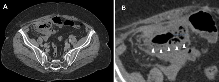
(A) Axial CT image at the level of the caecum and (B) magnified view of the caecum, demonstrating intramural gas (white arrowheads) and thickened bowel wall (blue dotted line).
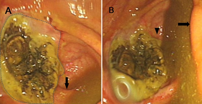
(A) Endoscopic view of the caecum, showing the area of ulcerated ischaemic mucosa (dashed outline), adjacent to this is the unaffected appendix orifice (black arrow); (B) endoscopic view of the caecum showing the ileocaecal valve (black arrow) and appendix orifice (black arrowhead).
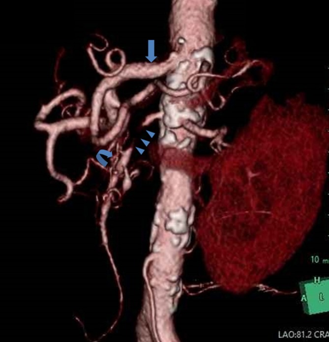
3D reconstruction of CT angiogram (left anterior oblique view), showing the coeliac axis (blue block arrow), tight SMA stenosis (blue arrowheads) and pancreatico-duodenal collaterals (curved arrow). SMA, superior mesenteric artery; 3D, three-dimensional.
References
-
- Montoro MA , Brandt LJ , Santolaria S , Gomollon F , Sánchez Puértolas B , Vera J , et al. . Clinical patterns and outcomes of ischaemic colitis: results of the Working Group for the Study of Ischaemic Colitis in Spain (CIE study) . Scand J Gastroenterol 2011. ; 46 : 236 – 46 . doi: 10.3109/00365521.2010.525794 - DOI - PubMed
LinkOut - more resources
Full Text Sources
