. 2019 Jul 16;19(4):e26.
doi: 10.4110/in.2019.19.e26. eCollection 2019 Aug.

Circulating CCR7loPD-1hi Follicular Helper T Cells Indicate Disease Activity and Glandular Inflammation in Patients with Primary Sjögren's Syndrome

Affiliations

Circulating CCR7loPD-1hi Follicular Helper T Cells Indicate Disease Activity and Glandular Inflammation in Patients with Primary Sjögren's Syndrome

Ji-Won Kim et al. Immune Netw. .

Abstract

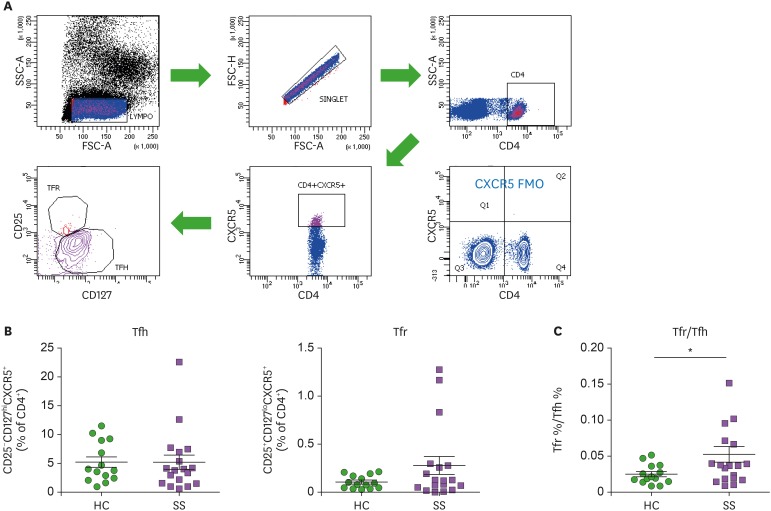
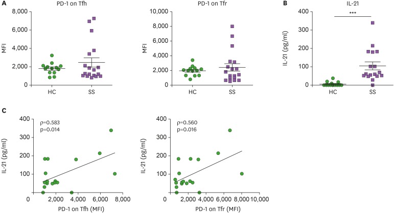
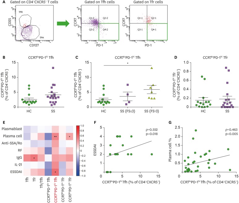
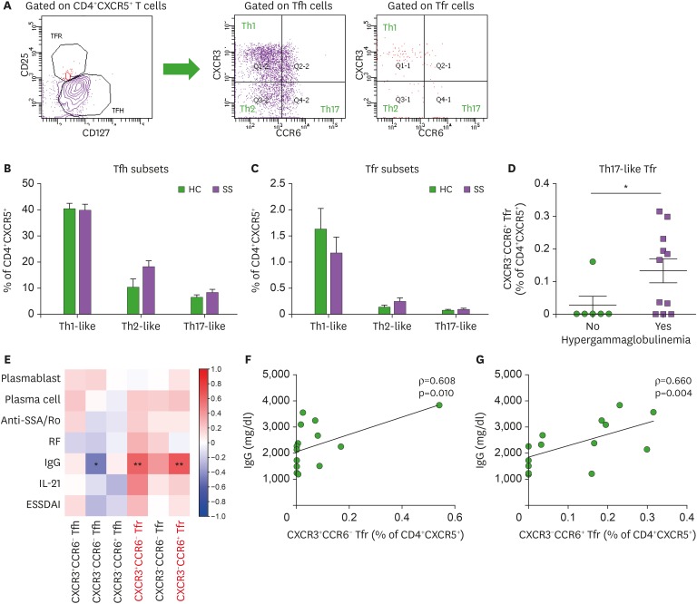
Since primary Sjögren's syndrome (pSS) is an autoummune disease of B cell hyperactivity and pathologic autoantibody response, follicular helper T (Tfh) cells and follicular regulatory T (Tfr) cells are suggested to be key players in pSS. We examined subsets of Tfh and Tfr cells from the blood in pSS patients, and whether these subsets represent disease activity, glandular inflammation, or autoantibody responses in pSS. Circulating Tfh and Tfr cells, along with their specific subsets, were identified from the peripheral blood of 18 pSS patients and 14 age- and sex-matched healthy controls (HCs) using flow cytometry analysis. Blood Tfr and Tfh cell ratios were increased in pSS patients compared with HCs. The CCR7loPD-1hi subset of circulating Tfh cells was increased in pSS patients with high degree of focal lymphocytic sialadenitis; whereas circulating Tfh cells did not differ between pSS patients and HCs. The frequency of CCR7loPD-1hi Tfh cells was significantly correlated with disease activity scores and differentiated B cells. PD-1 expression on blood Tfh and Tfr cells showed positive correlations with IL-21 in pSS. Increasing trend of blood Tfr cells was observed in pSS patients, and blood Tfr cells (particularly Th1 and Th17 subsets) represented hypergammaglobulinemia in pSS. In summary, circulating CCR7loPD-1hi Tfh cells indicated disease activity and glandular inflammation in pSS. Circulating Tfr cells, shifted toward Th1 and Th17 subsets, indicated ongoing IgG production in pSS. Subsets of circulating Tfh or Tfr cells could be biomarkers for disease monitoring and patient stratification in pSS.

Keywords: Autoantibodies; Sjögren's syndrome; T-lymphocyte subsets; T-lymphocytes; T-lymphocytes, regulatory.


Conflict of interest statement

Conflicts of Interest: The authors declare no potential conflicts of interest.

Figures

Figure 1
Figure 1. Circulating Tfh cells, Tfr cells, and Tfr/Tfh ratios in patients with pSS. (A) Gating strategies for circulating Tfh and Tfr cell populations representing CD25CD127hiCXCR5+CD4+ Tfh cells and CD25+CD127loCXCR5+CD4+ Tfr cells in the peripheral blood of pSS patients (n=18) and HCs (n=14). (B) Frequencies of Tfh and Tfr cells are compared between pSS patients and HCs. (C) The Tfr/Tfh ratio is increased in pSS patients compared to HCs. Bars indicate the means±SEMs.
*p<0.05.
Figure 2
Figure 2. PD-1 expression on Tfh and Tfr cells in peripheral blood is positively correlated with serum IL-21 levels in pSS. (A) PD-1 expression on circulating Tfh and Tfr cells from pSS patients (n=18) and HCs (n=14). (B) Serum IL-21 levels were increased in patients with pSS (n=17) as compared to HCs (n=14). (C) Correlation of intensity of PD-1 expression on Tfh and Tfr cells with serum IL-21 levels (n=17). Bars indicate the means±SEMs.
***p<0.001.
Figure 3
Figure 3. The CCR7loPD-1hi subset of circulating Tfh cells correlates with disease activity of pSS and represents glandular inflammation of pSS. (A) Gating strategies for CCR7loPD-1hi and CCR7hiPD-1lo subsets from circulating Tfh and Tfr cells. (B) Circulating CCR7loPD-1hi Tfh cells tended to increase in pSS patients (n=18) as compared to HCs (n=14), although circulating Tfh cells showed similar frequencies between pSS patients and HCs. (C) The CCR7loPD-1hi subset of circulating Tfh cells was markedly increased in pSS patients with FS >3 versus HCs (1-way ANOVA with post-hoc Bonferroni test). (D) Frequencies of circulating CCR7loPD-1hi Tfr cells are similar between pSS patients (n=18) and HCs (n=14). (E) Correlation plot of circulating Tfh and Tfr cells and their subsets with circulating plasmablast and plasma cell populations, serum anti-SSA/Ro Ab titers, serum RF titers, serum IgG levels, serum IL-21 levels, and the ESSDAI. (F) Correlation of circulating CCR7loPD-1hi Tfh cells with ESSDAI scores (n=18). (G) Correlation of circulating CCR7loPD-1hi Tfh cells with circulating plasma cells (n=32). Bars indicate the means±SEMs.
*p<0.05; **p<0.01.
Figure 4
Figure 4. Th1 and Th17 subsets of circulating Tfr cells indicate hypergammaglobulinemia in pSS. (A) Gating strategies for Th1-like, Th2-like, and Th17-like subsets of circulating Tfh and Tfr cells. (B and C) Th1-like, Th2-like, and Th17-like subsets of circulating Tfh and Tfr cells are compared between pSS patients (n=18) and HCs (n=13). Th1-like Tfh = CXCR3+CCR6 Tfh cells; Th2-like Tfh = CXCR3CCR6 Tfh cells; Th17-like Tfh = CXCR3-CCR6+ Tfh cells; Th1-like Tfr = CXCR3+CCR6- Tfr cells; Th2-like Tfr = CXCR3-CCR6- Tfr cells; Th17-like Tfr = CXCR3-CCR6+ Tfr cells. (D) Circulating CXCR3CCR6+ (Th17-like) Tfr cells in pSS patients with and without hypergammaglobulinemia. (E) Correlation plot of circulating Tfh and Tfr cell subsets with circulating plasmablast and plasma cell populations, serum anti-SSA/Ro Ab titers, serum RF titers, serum IgG levels, serum IL-21 levels, and the ESSDAI. (F) Correlation of circulating CXCR3+CCR6 (Th1-like) Tfr cells with serum IgG levels (n=17). (G) Correlation of circulating CXCR3-CCR6+ (Th17-like) Tfr cells with serum IgG levels (n=17). Bars indicate the means±SEMs.
*p<0.05; **p<0.01.

References

    1. Craft JE. Follicular helper T cells in immunity and systemic autoimmunity. Nat Rev Rheumatol. 2012;8:337–347. - PMC - PubMed
    1. Kwok SK, Lee J, Yu D, Kang KY, Cho ML, Kim HR, Ju JH, Lee SH, Park SH, Kim HY. A pathogenetic role for IL-21 in primary Sjögren syndrome. Nat Rev Rheumatol. 2015;11:368–374. - PubMed
    1. Jin L, Yu D, Li X, Yu N, Li X, Wang Y, Wang Y. CD4+CXCR5+ follicular helper T cells in salivary gland promote B cells maturation in patients with primary Sjögren's syndrome. Int J Clin Exp Pathol. 2014;7:1988–1996. - PMC - PubMed
    1. Kang KY, Kim HO, Kwok SK, Ju JH, Park KS, Sun DI, Jhun JY, Oh HJ, Park SH, Kim HY. Impact of interleukin-21 in the pathogenesis of primary Sjögren's syndrome: increased serum levels of interleukin-21 and its expression in the labial salivary glands. Arthritis Res Ther. 2011;13:R179. - PMC - PubMed
    1. Gong YZ, Nititham J, Taylor K, Miceli-Richard C, Sordet C, Wachsmann D, Bahram S, Georgel P, Criswell LA, Sibilia J, et al. Differentiation of follicular helper T cells by salivary gland epithelial cells in primary Sjögren's syndrome. J Autoimmun. 2014;51:57–66. - PubMed