Review
. 2020 Feb;77(4):573-591.
doi: 10.1007/s00018-019-03285-0. Epub 2019 Sep 9.

Regulation of gap junction intercellular communication by connexin ubiquitination: physiological and pathophysiological implications

Affiliations
Review

Regulation of gap junction intercellular communication by connexin ubiquitination: physiological and pathophysiological implications

Max Zachrisson Totland et al. Cell Mol Life Sci. 2020 Feb.

Abstract

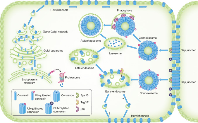
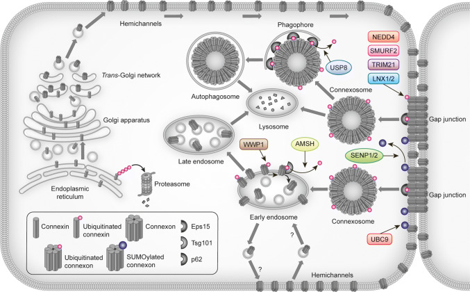
Gap junctions consist of arrays of intercellular channels that enable adjacent cells to communicate both electrically and metabolically. Gap junctions have a wide diversity of physiological functions, playing critical roles in both excitable and non-excitable tissues. Gap junction channels are formed by integral membrane proteins called connexins. Inherited or acquired alterations in connexins are associated with numerous diseases, including heart failure, neuropathologies, deafness, skin disorders, cataracts and cancer. Gap junctions are highly dynamic structures and by modulating the turnover rate of connexins, cells can rapidly alter the number of gap junction channels at the plasma membrane in response to extracellular or intracellular cues. Increasing evidence suggests that ubiquitination has important roles in the regulation of endoplasmic reticulum-associated degradation of connexins as well as in the modulation of gap junction endocytosis and post-endocytic sorting of connexins to lysosomes. In recent years, researchers have also started to provide insights into the physiological roles of connexin ubiquitination in specific tissue types. This review provides an overview of the advances made in understanding the roles of connexin ubiquitination in the regulation of gap junction intercellular communication and discusses the emerging physiological and pathophysiological implications of these processes.

Keywords: Cancer; Cataract; Electrical synapse; Heart arrhythmia; Heart ischemia; Lens; NEDD4; Ubiquitin.


Figures

Fig. 1
Fig. 1
Connexins, connexons and gap junction channels. a Connexins have four transmembrane domains, which are connected by two extracellular loops and one intracellular loop. The N- and C-termini are both located in the cytosol. b Connexins form hexamers called connexons. Connexins can combine with either the same or different connexin isoforms, forming homomeric or heteromeric connexons, respectively. c Connexons form gap junction channels by interacting with either identical homomeric or heteromeric connexons in adjacent cells, forming homotypic channels, or with different homomeric or heteromeric connexons, forming heterotypic channels
Fig. 2
Fig. 2
Intracellular trafficking and ubiquitination and SUMOylation of connexins. Connexins are co-translationally inserted into the endoplasmic reticulum and transported through the Golgi, through the trans-Golgi and to the plasma membrane. Along their trafficking to the plasma membrane, connexins oligomerize into connexons. A subpool of the newly synthesized connexins undergoes ERAD, in which connexins are retrotranslocated to the cytosol and degraded by proteasomes. In the plasma membrane, connexons can function as hemichannels or diffuse to the periphery of gap junctions, where they can dock with connexons in adjacent cells to form intercellular channels. During endocytosis of gap junctions, both membranes of the junctions are internalized into one of the cells, thereby forming a connexosome, also called an annular gap junction. Three different pathways for trafficking of connexins to lysosomes are illustrated in the figure: (1) direct fusion between connexosomes and lysosomes, (2) sequestration of the connexosome by a phagophore and subsequent fusion between an autophagosome containing the connexosome and a lysosome, and (3) transformation of the connexosome into a connexin-enriched, multivesicular endosome with a single limiting membrane, which is associated with the fusion between the connexosome and early endosomes. Connexins are then sorted from early endosomes via late endosomes to lysosomes. Connexons at the plasma membrane that are not assembled into gap junctions can also undergo endocytosis, but their intracellular trafficking is poorly characterized. They are possibly transported to early endosomes (indicated by a question mark). Following endocytosis, connexins may undergo recycling to the plasma membrane, possibly from the early endosomes (indicated by a question mark). Connexin ubiquitination has been suggested to be involved in ERAD of certain connexin isoforms, gap junction endocytosis, autophagy-mediated degradation of connexins and sorting of connexins from early endosomes to lysosomes. Eps15 has been suggested to bind to ubiquitinated Cx43 at the plasma membrane and to control gap junction endocytosis and autophagy-mediated degradation. Tsg101 is part of ESCRT and has been suggested to bind to ubiquitinated Cx43 at the limiting membrane of early endosomes and to regulate its sorting into the lumen of the endosome. p62 has been suggested to bind to ubiquitinated Cx43 after gap junction endocytosis and to be involved in the sequestration of connexosomes by autophagosomes. Cx43 has also been shown to be modified by SUMOylation, which has been suggested to stabilize Cx43 and consequently cause increased gap junction size
Fig. 3
Fig. 3
Overview of proteins involved in controlling gap junctions by connexin ubiquitination or SUMOylation. The figure depicts the proteins identified to date that participate in the regulation of gap junction intercellular communication through connexin ubiquitination or SUMOylation and the possible subcellular localizations where they may interact with connexins. For details, see Table 2 and the main text

References

    1. Saez JC, Berthoud VM, Branes MC, Martinez AD, Beyer EC. Plasma membrane channels formed by connexins: their regulation and functions. Physiol Rev. 2003;83:1359–1400. - PubMed
    1. Beyer EC, Berthoud VM. Gap junction gene and protein families: connexins, innexins, and pannexins. Biochim Biophys Acta. 1860;2018:5–8. - PMC - PubMed
    1. Goodenough DA, Paul DL. Gap junctions. Cold Spring Harb Perspect Biol. 2009;1:a002576. - PMC - PubMed
    1. Aasen T. Connexins: junctional and non-junctional modulators of proliferation. Cell Tissue Res. 2015;360:685–699. - PubMed
    1. Vinken M. Introduction: connexins, pannexins and their channels as gatekeepers of organ physiology. Cell Mol Life Sci. 2015;72:2775–2778. - PMC - PubMed

LinkOut - more resources