. 2019 Nov;302(11):1958-1967.
doi: 10.1002/ar.24251. Epub 2019 Oct 1.

High-Resolution Computed Tomography Imaging of the Cranial Arterial System and Rete Mirabile of the Cat (Felis catus)

Affiliations

High-Resolution Computed Tomography Imaging of the Cranial Arterial System and Rete Mirabile of the Cat (Felis catus)

E Leon Kier et al. Anat Rec (Hoboken). 2019 Nov.

Abstract

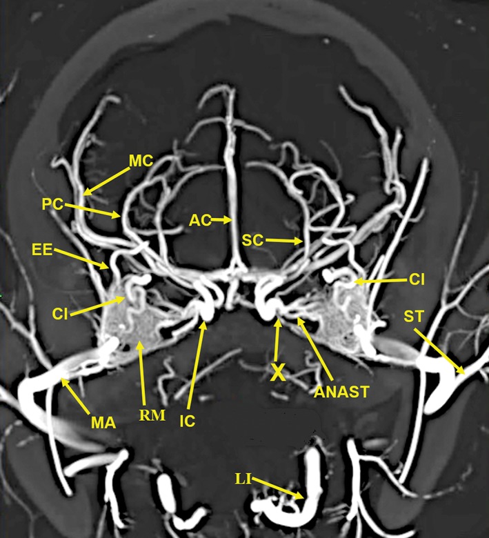
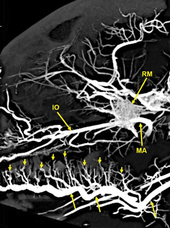
The objective of this study was to investigate the possibility of obtaining high-resolution multiplanar computed tomography (CT) imaging of the cranial arterial circulation of the cat (Felis catus), the rete mirabile, and components of the skull, utilizing preserved cat specimens with an arterial system that was injected with a radiopaque contrast compound in the early 1970s. Review of the literature shows no high-resolution CT studies of the cat's cranial circulation, with only few plain radiographic studies, all with limited cranial vascular visualization. In view of the inability of the radiographic techniques available from 1970s to mid-2000s to provide high-resolution imaging of the arterial circulation within the intact skull and brain of the cat, without dissection and histologic sectioning and disruption of tissues, no further imaging was performed for many years. In 2010, a high-resolution micro CT scanner became available, large enough to scan the entire nondissected head of the arterially injected cats. All the obtained CT images were processed with a software program that provided 3D volume rendering and multiplanar reconstruction with the ability to change the plane angulation and slab thickness. These technical features permitted more precise identification of specific arterial and bony anatomy. The obtained images demonstrated, with a nondestructive method, high-resolution vascular anatomy of the cerebral, orbital, facial arterial system, the rete mirabile, and skull bone components of the cat, with details not previously described in the literature. Anat Rec, 302:1958-1967, 2019. © 2019 The Authors. The Anatomical Record published by Wiley Periodicals, Inc. on behalf of American Association of Anatomists.

Keywords: Felis catus; carotid rete; cat; cerebral arteries; extracranial rete; facial arteries; infraorbital canal; lacrimal canal; multiplanar high-resolution micro-computed tomography (CT); orbital arteries; rete mirabile; skull bones; sphenopalatine foramen.


Conflict of interest statement

The authors declare no competing or financial interest.

Figures

Figure 1
Figure 1
Lateral (A) and dorsoventral (B) 3D volume rendered projections of the cranial arterial circulation of a cat. Of note is the extent of the demonstrated vasculature in the bone and soft tissue free images and the small size of the vasculature when compared with the yellow colored 1.0‐mm scale bar (arrow). These 3D images can be rotated and turned in any direction or angle needed for the identification of a specific blood vessel.
Figure 2
Figure 2
A 25‐mm‐thick lateral multiplanar reconstruction of the cranial arterial circulation of the cat, using maximum intensity projection. A large number of cranial arteries can be identified. The anastomotic blood vessels (ANAST) extending from the rete mirabile (RM) to the reformed internal carotid artery (IC) are demonstrated. The stump of the origin of the vestigial internal carotid artery (IC‐S) is identified. The tortuous course of the ciliary artery (CI) within the external rete is demonstrated. The position of the maxillary artery (MA) is in the inferior section of the rete mirabile (RM). Arterial abbreviations: AC, anterior cerebral; AP, ascending pharyngeal; BA, basilar; CC, common carotid; EC, external carotid; EE, external ethmoid; FA, facial; GP, greater palatine; IA, inferior alveolar; IC, internal carotid; IE, internal ethmoid; IO, infraorbital; LI, lingual; MC, middle cerebral; OC, occipital; PA, posterior auricular; PC, posterior cerebral; SC, superior cerebellar; ST, superficial temporal; VA, vertebral.
Figure 3
Figure 3
A 20‐mm‐thick oblique dorsoventral multiplanar reconstruction of the cranial arterial circulation of the cat, using maximum intensity projection. Among the many identified vascular structures are (1) The rete mirabile (RM). (2) The junction (X) of the multiple anastomotic vessels (ANAST) from the medial edge of the rete mirabile (RM), forming the internal carotid artery (IC). (3) The circle of Willis formed by internal carotid (IC), posterior cerebral/posterior communicating arteries (PC), and a small segment of the anterior cerebral (AC) arteries. (4) The origin of the external ethmoidal (EE) arteries within the rete (RM) is demonstrated. (5) The tortuous course of the ciliary artery (CI) within the rete (RM) also is demonstrated. (6) The long posterior ciliary artery (LPC) and the faint outline of the ciliary circle branches (small arrows) can be identified. (7) The position of the maxillary artery (MA) is in the inferolateral section of the rete mirabile (RM). Arterial abbreviations: AIC, anterior inferior; AP, ascending pharyngeal; BA, basilar; CH, chiasmatic; EC, external carotid; FA, facial; IE, internal ethmoid; LA, lacrimal; LI, lingual; PA, posterior auricular; PIC, posterior inferior; SC, superior cerebellar; ST, superficial temporal; VE, vertebral.
Figure 4
Figure 4
A 10‐mm‐thick oblique dorsoventral multiplanar reconstruction of the cranial arterial circulation of the cat, using maximum intensity projection. This thin slab shows the maxillary artery (MA) coursing within the rete (RM). The anastomotic blood vessels (ANAST) originating from the medial edge of the rete mirabile are coursing toward the origin of the reconstituted and functional internal carotid artery (IC). The ascending pharyngeal artery (AP) and the middle meningeal artery (MM) also have an important role in forming the reconstituted internal carotid artery. The region (X) is where the anastomotic blood vessels from the external rete, the ascending pharyngeal artery (AP), and the middle meningeal artery join to form the reconstituted functional internal carotid artery (IC). Arterial abbreviation: EC, external carotid artery.
Figure 5
Figure 5
A 20‐mm‐thick anteroposterior multiplanar reconstruction of the cranial arterial circulation of the cat, using maximum intensity projection. Of note is the junction (X) of the multiple anastomotic vessels (ANAST) arising from the medial edge of the rete mirabile (RM), forming the internal carotid artery (IC). Also visible are the origin of the external ethmoid (EE) within the rete. The ciliary artery (CI), which has a tortuous course within the rete, is actually a branch of the internal maxillary artery (IM). Arterial abbreviations: AC, anterior cerebral; LI, lingual; MC, middle cerebral; PC, posterior cerebral; SC, superior cerebellar; ST, superficial temporal.
Figure 6
Figure 6
A 20‐mm‐thick lateral multiplanar reconstruction of the cranial arterial circulation of another cat, using maximum intensity projection. A large number of very small arteriolar branches (small arrows) of the lingual artery (longer arrows) are penetrating the tongue. Arterial abbreviations: IO, infraorbital; MA, maxillary; RM, rete mirabile.
Figure 7
Figure 7
A 13‐mm‐thick dorsoventral multiplanar reconstruction of the cranium of the arterially injected cat using maximum intensity projection. The skull components are orange colored annotations; the arterial structures are in yellow color annotation in Figures 7, 8, 9, 10, 11, 12. The nasal cavity (NC) walls are the maxilla (MAX), ethmoid (ET), and orbitosphenoid (OSP) bones. The rete mirabile (RM) and its component, the tortuous ciliary artery (CL), pass through the orbital fissure, and the alisphenoid (ASP). Also identified are segments of the following components of the skull: C1, posterior arch of first cervical vertebra; CP, coronoid process; OC, occipital; POP/ZA, post orbital process of the zygomatic arch; PSP, presphenoid; TB, tympanic bulla; ZA, zygomatic arch; ZY, zygoma; and the following arteries: AC, anterior cerebral; BA, basilar; IC, internal carotid; IE, internal ethmoid; MC, middle cerebral; PC, posterior cerebral; SC, superior cerebellar.
Figure 8
Figure 8
A 6‐mm‐thick anteroposterior multiplanar reconstruction of the cranium of the arterially injected cat using maximum intensity projection. The middle cerebral (MC) artery branches, the anterior cerebral (AC) artery branches, within the interhemispheric fissure, and the lenticulostriate (LS) arteries of the cerebrum are encased superiorly and laterally by the parietal (PA) bones and inferiorly by the alisphenoid (ASP) bones. The rete mirabile (RM) is situated lateral to the pterygoid bone (PT) with its inferior extension; the hamulus (HA). The pterygoid bone is fused medially with the basisphenoid (BS). Anastomotic (ANAST) arterial branches extend medially to participate in the reconstitution of the internal carotid artery at the circle of Willis as seen in Figure 5. Segments of the zygomatic arch (ZY) and of the mandibular ramus (MR) including the inferior alveolar artery (IA) are demonstrated.
Figure 9
Figure 9
An 11‐mm‐thick dorsoventral multiplanar reconstruction of the cranium of the arterially injected cat using maximum intensity projection. The sphenopalatine (SP) arteries pass through the sphenopalatine foramina (SPF) to enter the nasal cavities. The sphenopalatine foramen is within the orbital process of the palatine (PA) bone. The vascular pattern in the nasal cavity is demonstrated in Figure 10. The sphenopalatine arteries are branches of the infraorbital (IO) arteries, which are the continuation of the maxillary (MA) arteries. The infraorbital artery continues anteriorly to pass through the infraorbital canal (IOC), which is within the zygoma (ZY) at the junction with the maxilla (MAX). The entire infraorbital canal with the infraorbital artery within it is seen in Figure 10. Also identified are segments of the bony basisphenoid (BS), the pterygoid (PT) bone, and the coronoid process (CP) of the mandible.
Figure 10
Figure 10
An 8‐mm‐thick dorsoventral multiplanar reconstruction of the cranium of the arterially injected cat using maximum intensity projection. The infraorbital artery (IO) passes through the infraorbital canal (IOC), within the zygoma (ZY). The latter is attached to the maxilla (MAX), forming the wall of the nasal cavity. The turbinates (TU) within the nasal cavity are supplied by branches of the sphenopalatine (SP) arteries which also surround the nasal septum (NS). The palatine (PA) bone adjacent to the sphenopalatine artery is identified. The inferior alveolar artery (IA) is within the mandibular canal (MAC) in the left mandibular ramus (MR) and in the mandibular foramen (MF) of the right mandibular ramus.
Figure 11
Figure 11
An 11‐mm‐thick anteroposterior multiplanar reconstruction of the cranium of the arterially injected cat using maximum intensity projection. The nasal (NA) bones form the roof of the nasal cavity. The bony nasal septum (NS) is attached at the suture between the two nasal bones. The lateral wall of the nasal cavity is formed by the maxilla (MAX), the lacrimal (LA) bone, and the segment of the zygoma (ZY) medial to the infraorbital canal (IOC). There is an extensive number of turbinates (TU) within the nasal cavity which are supplied by branches of the sphenopalatine (SP) arteries. The infraorbital artery (IO) passing through the infraorbital canal is identified with the smaller lacrimal canal (LC), for the lacrimal duct, adjacent to the infraorbital canal. The bony palate, the floor of the nasal cavity, at this level is formed laterally by the maxilla (MAX) and medially by the palatine (PA) bone. Also identified are the anterior palatine arteries (AP) below the palate; the inferior alveolar artery (IA) within the mandibular canal (MAC) in the mandibular body (MB) and the upper (UPM) and lower (LPM) premolar teeth.
Figure 12
Figure 12
An 11‐mm‐thick dorsoventral multiplanar reconstruction of the mandible of the arterially injected cat using maximum intensity projection. Visualized are inferior alveolar (IA) arteries within the mandibular canals (MC) of the mandible (MB). The inferior alveolar arteries exit the mental foramina (MEF) becoming the mental (ME) arteries. The mandibular symphysis (MS) is visible.

Similar articles

Cited by

References

    1. Baker MA. 1972. Influence of the carotid rete on brain temperature in cats exposed to hot environments. J Physiol 220:711–728. - PMC - PubMed
    1. du Boulay G, Darling M. 1975. Autoregulation in external carotid artery branches in the baboon. Neuroradiology 9:129–132. - PubMed
    1. du Boulay GH, Verity PM. 1973. The cranial arteries of mammals. London: W. Heinemann.
    1. du Boulay G, Kendall BE, Crockard A, Sage M, Belloni G. 1975. The autoregulatory capability of Galen's rete cerebri and its connections. Neuroradiology 9:171–181. - PubMed
    1. Chiasson RB, Booth ES. 1973. Laboratory anatomy of the cat. Dubuque, Iowa: WM. C. Brown Company.

MeSH terms