. 2019 Sep 10;11(17):7242-7256.
doi: 10.18632/aging.102259. Epub 2019 Sep 10.

Reactive oxygen species-mediated senescence is accelerated by inhibiting Cdk2 in Idh2-deficient conditions

Affiliations

Reactive oxygen species-mediated senescence is accelerated by inhibiting Cdk2 in Idh2-deficient conditions

Unbin Chae et al. Aging (Albany NY). .

Abstract

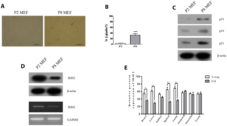
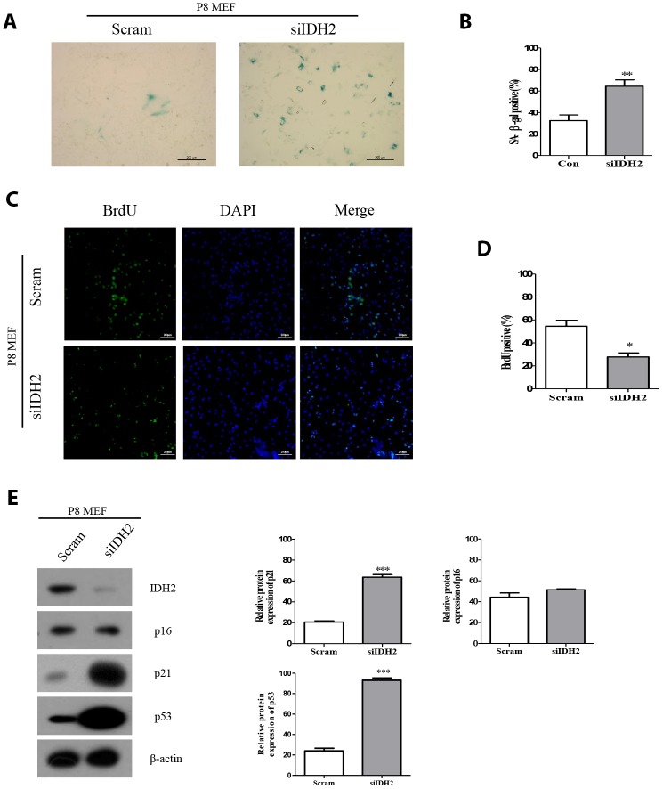
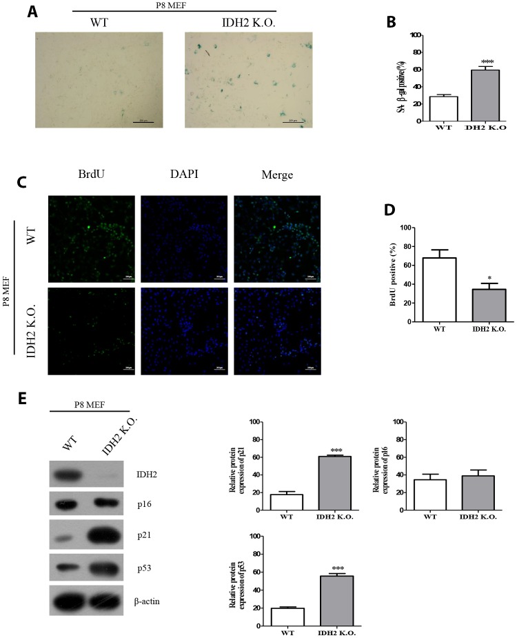
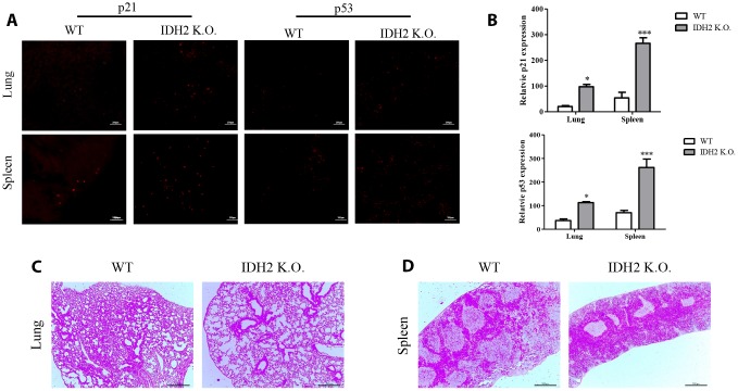
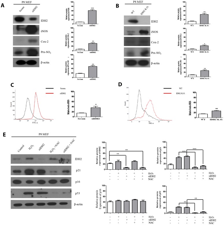
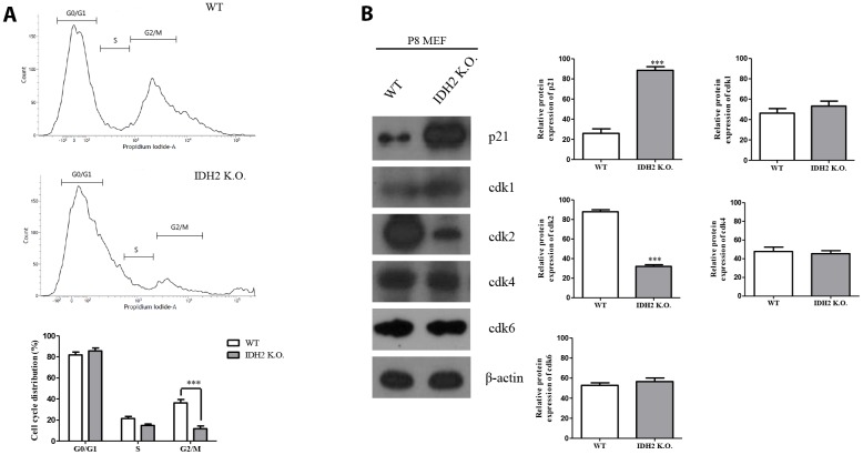
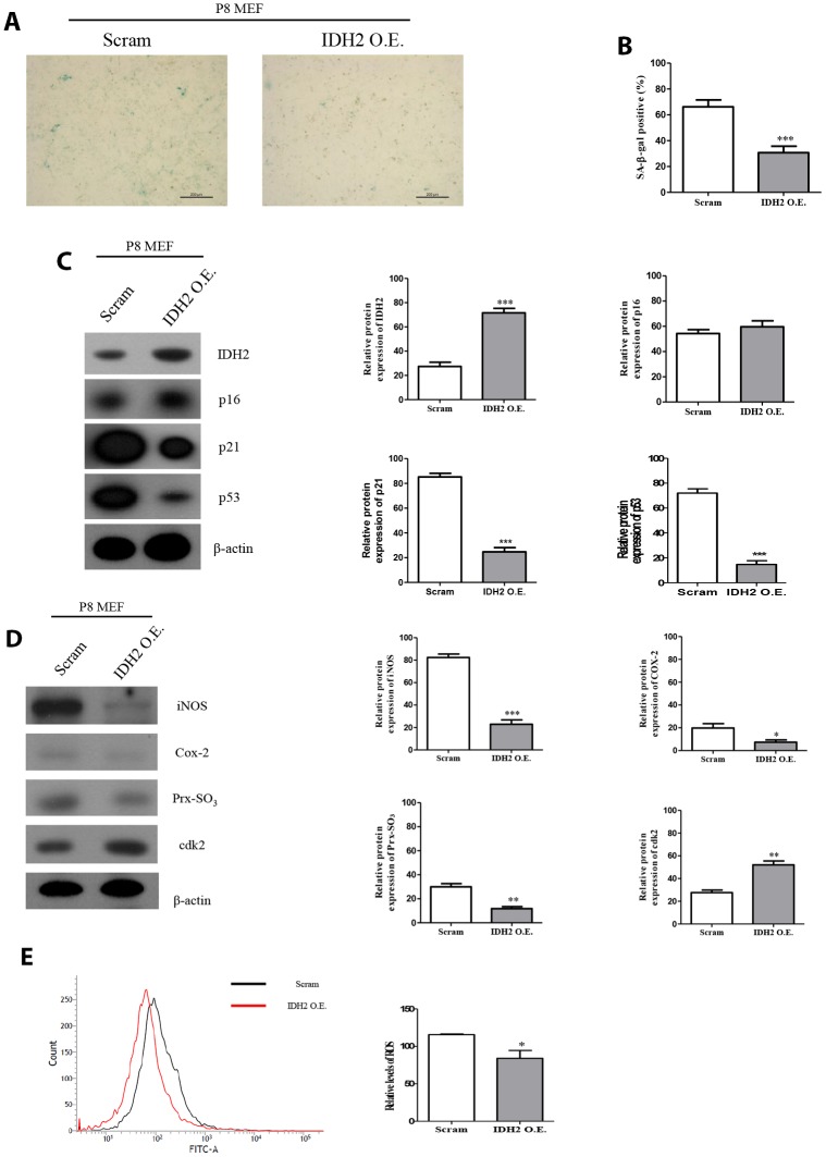
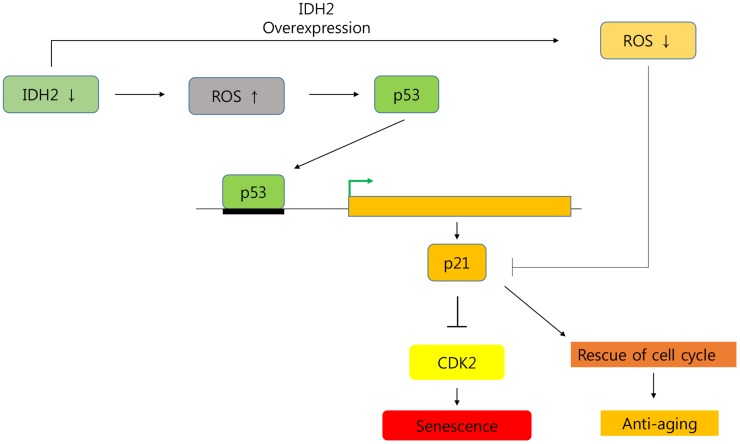
Among the many factors that promote cellular senescence, reactive oxygen species (ROS) are a focus of intense research because of their critical role in accelerating cellular senescence and initiating senescence-related diseases that can be fatal. Therefore, maintaining the proper balance of ROS in cells is a key method to alleviate senescence. Recent studies have found that isocitrate dehydrogenase 2 (IDH2), a critical enzyme of the tricarboxylic acid cycle, participates in ROS generation and in cellular dysfunction that is induced by excessive levels of ROS. Loss of IDH2 induces mitochondrial dysfunction that promotes excessive ROS generation and the development of several diseases. The results of this study suggest that Idh2 plays an important role in cellular senescence. Idh2 deficiency resulted in senescence-associated phenotypes and increased levels of senescence marker proteins in mouse embryonic fibroblasts and tissues. Furthermore, excessive ROS were generated in Idh2-deficient conditions, promoting cellular senescence by inducing cell cycle arrest through cyclin-dependent kinase 2. These results indicate that loss of Idh2 is a critical factor in regulating cellular senescence. Taken together, our findings contribute to the field of senescence research and suggest that IDH2 is a potential target of future anti-senescence studies.

Keywords: cell cycle; cyclin-dependent kinase 2 (Cdk2); isocitrate dehydrogenase 2 (IDH2); reactive oxygen species (ROS); senescence.


Conflict of interest statement

CONFLICTS OF INTEREST: The authors have declared that no competing interests exist.

Figures

Figure 1
Figure 1
Idh2 was downregulated in senescent mouse embryonic fibroblasts (MEFs) and aging tissues. (A) SA-ß-gal staining of Passage 2 (P2) and Passage 8 (P8) MEFs. Scale bar, 200 µm. (B) Statistical analysis of SA-ß-gal-stained positive cells between P2 and P8 MEFs. (C) Western blot analysis between P2 and P8 MEFs. The following antibodies were used: anti-p53, anti-p16, anti-p21, and anti-ß-actin. (D) Detection of Idh2 expression level in P2 and P8 MEFs. Western blotting and reverse-transcriptase PCR was performed for detecting Idh2. (E)Relative protein expression of Idh2 was detected in mouse tissues using western blot analysis. Ten-week-old and 47-week-old mice were used. Data are expressed as means ± SD (n = 3). *p < 0.05, **p < 0.01, and ***p < 0.001.
Figure 2
Figure 2
Downregulation of Idh2 in senescent mouse embryonic fibroblasts (MEFs) accelerates the senescence phenotype. (A) SA-β-gal staining of control and Idh2-silenced MEFs. Passage 8 (P8) MEFs were used. Scale bar, 200 μm. (B) Statistical analysis of SA-β-gal-stained positive cells between control and Idh2-knockdown MEFs. (C) BrdU levels between control and Idh2-knockdown MEFs as determined by immunocytochemistry. Nuclei were stained with DAPI, and the merged images show BrdU and DAPI signals. Scale bar, 10 μm. (D) Statistical analysis of BrdU-positive cells between control and Idh2-knockdown MEFs. (E) Western blot analysis between control and Idh2-knockdown MEFs. The following antibodies were used for detection: anti-Idh2, anti-p16, anti-p21, anti-p53, and anti- β-actin. Data are expressed as means ± SD (n = 3). *p < 0.05, **p < 0.01, and ***p < 0.001.
Figure 3
Figure 3
The senescence phenotype is promoted in Idh2 knockout mouse embryonic fibroblasts (MEFs). (A) SA-β-gal staining of wild type and Idh2 knockout MEFs. Passage 8 (P8) MEFs were used. Scale bar, 200 μm. (B) Statistical analysis of SA-β-gal-stained positive cells between wild type and Idh2 knockout MEFs. (C) BrdU level between wild type and Idh2 knockout MEFs as determined by immunocytochemistry. Nuclei were stained with DAPI, and the merged images show BrdU and DAPI signals. Scale bar, 10 μm. (D) Statistical analysis of BrdU-positive cells between wild type and Idh2 knockout MEFs. (E) Western blot analysis between wild type and Idh2 knockout MEFs. The following antibodies were used for detection: anti-Idh2, anti-p16, anti-p21, anti-p53, and anti-β-actin. Data are expressed as means ± SD (n = 3). *p < 0.05, **p < 0.01, and ***p < 0.001.
Figure 4
Figure 4
Acceleration of senescence in several tissues is observed in Idh2 knockout mice. (A) P21 and p53 levels in lung and spleen tissues from wild type and Idh2 knockout mice as determined by immunohistochemistry. Images show p21 and p53 signals. Scale bar, 10 μm (n = 6/group). (B) Statistical analysis of p21 and p53-positive signals between lung and spleen tissues from wild type and Idh2 knockout mice. (C) Representative H&E-stained sections from wild type and Idh2 knockout mouse lungs (n = 6/group). (D) Representative H&E-stained sections from wild type and Idh2 knockout mouse spleens (n = 6/group).
Figure 5
Figure 5
Idh2 deficiency-mediated reactive oxygen species (ROS) generation activates p21 expression. (A) Western blot analysis of pro-inflammatory mediators and oxidative stress marker proteins in control and Idh2-knockdown mouse embryonic fibroblasts (MEFs). The following antibodies were used for detection: anti-Idh2, anti-iNOS, anti-Cox-2, anti-Prx-SO3, and anti-β-actin. (B) Western blot analysis of pro-inflammatory mediators and oxidative stress marker proteins between wild type and Idh2 knockout MEFs. The following antibodies were used for detection: anti-Idh2, anti-iNOS, anti-Cox-2, anti-Prx-SO3, and anti-β-actin. (C) Control and Idh2-knockdown MEFs were incubated with DCF-DA for 15 min at 37°C and intracellular ROS levels were analyzed by flow cytometry. (D) Wild type and Idh2 knockout MEFs were incubated with DCF-DA for 15 min at 37°C and intracellular ROS levels were analyzed by flow cytometry. (E) After transfecting MEFs with siIdh2, H2O2 was added for 3 days. NAC was added 4 h before H2O2 treatment. Western blot analysis was detected with the following antibodies. Data are expressed as means ± SD (n = 3). *p < 0.05, **p < 0.01, and ***p < 0.001.
Figure 6
Figure 6
Downregulation of Idh2 promotes senescence in mouse embryonic fibroblasts (MEFs) through suppressing Cdk2. (A) Wild type and Idh2 knockout MEFs were incubated with propidium iodide for 15 min at 37°C. Cell cycle was detected with flow cytometry. (B) Cyclin-dependent kinase protein levels were detected with western blot analysis in wild type and Idh2 knockout MEFs. The following antibodies were used for detection: anti-p21, anti-Cdk1, anti-Cdk2, anti-Cdk4, anti-Cdk6, and anti-β-actin. Data are expressed as means ± SD (n = 3). *p < 0.05, **p < 0.01, and ***p < 0.001.
Figure 7
Figure 7
Overexpression of Idh2 prevents the acceleration of senescence in mouse embryonic fibroblasts (MEFs). (A) SA-β-gal staining of control and Idh2-overexpressing MEFs. pLenti 6.3 Idh2 plasmid was transfected into Passage 8 (P8) MEFs. (B) Statistical analysis of SA-β-gal-stained positive cells between control and Idh2-overexpressing MEFs. (C) Senescence-associated marker proteins were detected using western blot analysis in control and Idh2-overexpressing MEFs. The following antibodies were used for detection: anti-Idh2, anti-p16, anti-p21, anti-p53, and anti-β-actin. (D) Pro-inflammatory mediators, reactive oxygen species (ROS) marker proteins, and cyclin-dependent kinase 2 were detected using western blot analysis. The following antibodies were used for detection: anti-iNOS, anti-Cox-2, anti-Prx-SO3, and anti-β-actin. (E) Relative intracellular ROS levels were detected in control and Idh2-overexpressing MEFs. Intracellular ROS were detected by flow cytometry. Data are expressed as means ± SD (n = 3). *p < 0.05, **p < 0.01, and ***p < 0.001.
Figure 8
Figure 8
Graphical abstract of the senescence pathway in Idh2-deficient conditions. Idh2 deficiency-induced ROS generation accelerates p53 and p21 signaling pathways which inhibit Cdk2 expression. Furthermore, overexpression of Idh2 prevented senescence-associated phenotypes by decreasing the levels of senescence signaling pathway-associated proteins p21 and p53.

Similar articles

Cited by

References

    1. LeBrasseur NK, Tchkonia T, Kirkland JL. Cellular Senescence and the Biology of Aging, Disease, and Frailty. Nestle Nutr Inst Workshop Ser. 2015; 83:11–18. 10.1159/000382054 - DOI - PMC - PubMed
    1. Cui H, Kong Y, Zhang H. Oxidative stress, mitochondrial dysfunction, and aging. J Signal Transduct. 2012; 2012:646354. 10.1155/2012/646354 - DOI - PMC - PubMed
    1. Shaw PX, Werstuck G, Chen Y. Oxidative stress and aging diseases. Oxid Med Cell Longev. 2014; 2014:569146. 10.1155/2014/569146 - DOI - PMC - PubMed
    1. Zorov DB, Juhaszova M, Sollott SJ. Mitochondrial reactive oxygen species (ROS) and ROS-induced ROS release. Physiol Rev. 2014; 94:909–50. 10.1152/physrev.00026.2013 - DOI - PMC - PubMed
    1. Shadel GS, Horvath TL. Mitochondrial ROS signaling in organismal homeostasis. Cell. 2015; 163:560–69. 10.1016/j.cell.2015.10.001 - DOI - PMC - PubMed

Publication types

Substances

LinkOut - more resources