Case Reports
. 2019 Nov;7(11):e923.
doi: 10.1002/mgg3.923. Epub 2019 Sep 10.

A rare case of acquired immunodeficiency associated with myelodysplastic syndrome

Affiliations
Case Reports

A rare case of acquired immunodeficiency associated with myelodysplastic syndrome

Juanjuan Li et al. Mol Genet Genomic Med. 2019 Nov.

Abstract

Background: Pediatric myelodysplastic syndromes (MDS) display clonal genomic instability that can lead to acquisition of other hematological disorders, usually by loss of heterozygosity. Immunodeficiency caused by uniparental disomy (UPD) has not previously been reported.

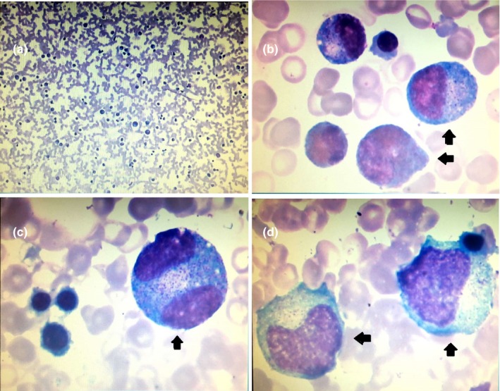
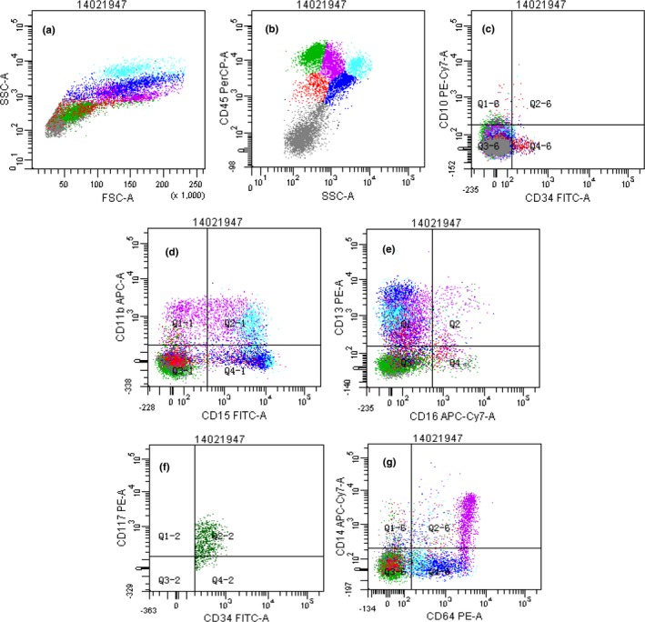
Methods: We investigated a 13-year-old boy who suffered from recurrent infections and pancytopenia for 1 year. Both the comet assay and chromosome breakage analysis were normal, but the bone marrow showed evidence of dysplasia characteristic of MDS. With his normal sister as donor, he underwent failed hematopoietic stem cell transplantation (HSCT) with reduced intensity conditioning (RIC) followed by successful HSCT with myeloablative conditioning (MAC). We used single nucleotide polymorphism (SNP) array, targeted gene panel, and whole exome sequencing to investigate the etiology of his disease.

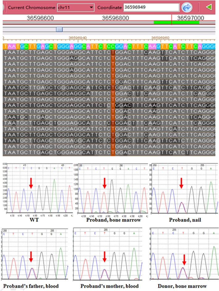
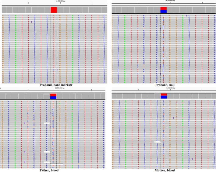
Results: The molecular analyses revealed multiple regions of homozygosity, one region encompassing a homozygous missense variant of recombination activating gene 1 (RAG1) which was previously associated with severe immunodeficiency in infancy. This RAG1 mutation was heterozygous in the proband's fingernail DNA, but was changed to homozygous in the proband's marrow by somatic acquisition of UPD event. No other pathogenic driver mutation for MDS-related genes was identified.

Conclusion: The hematological phenotype, somatic genomic instability, and response to HSCT MAC but not HSCT RIC deduced to a diagnosis of MDS type refractory cytopenia of children in this patient. His immunodeficiency was secondary to MDS due to somatic acquisition of homozygosity for known pathogenic RAG1 mutation.

Keywords: RAG1; acquired UPD; immunodeficiency; myelodysplastic syndrome.


Conflict of interest statement

The authors declare that there is no conflict of interest.

Figures

Figure 1
Figure 1
Bone marrow aspiration smear of the patient (Wright‐Giemsa stain). Panel a shows granulocyte series hypoplasia with slightly increasing proportion of myeloblasts and neutrophilic promyelocyte and scarity of bands and segmented neutrophils. Panels b, c and d show abnormal promyelocyte with immature nuclear and aging cytoplasm (arrowhead). The promyelocyte in Panel c is binuclear (arrowhead). Bone marrow aspiration smear showed the percentages of different lineage: 2% myeloblasts, 5% promyelocytes, 6.5% myelocytes, 1.5% metamyelocytes, 0.5% bands, 1% segmented neutrophils, 47.5% lymphocytes, 0.5% normoblasts, 6% rubricytes, 22.5% metarubricytes, and 2.5% monocytes
Figure 2
Figure 2
Immunophenotyping of patient’s bone marrow sample by flow cytometry. Panel a shows a global overview of bone marrow cellular compartments projected on SSC graphs, including lymphocyte (green, 25.0%), monocyte (purple, 15.1%), granulocyte (blue, 14.4%), eosinophil (light blue, 8.0%), blast cell or pre‐B cell (red, 4.7%) and erythroblast (gray, 32.8%). The myeloid cells had reduced CD45 and SSC expression (Panel b), and also lacked the expression of CD10, CD11b and CD16 (Panels c, d, e). The myeloid cells expressing CD13+/CD16+ showed an abnormal distribution pattern, indicating irregular myeloid differentiation (Panel e). The myeloblasts with expression of CD34 and CD117 (CD34+/CD117+) accounted for approximate 1% of all the nucleated cells observed, indicating a slight increase of the ratio (Panel f). The monocytes accounted for approximate 15.1% of all the nucleated cells indicative of an increase of the ratio and did not express significant abnormality (Panel g). The fluorochrome‐conjugated monoclonal antibodies (PerCP, FITC, PE‐Cy7, PE, APC‐Cy7 and APC) were used to stain the following antigens: HLA‐DR, CD2, CD3, CD4, CD5, CD7, CD8, CD10, CD11b, CD13, CD14, CD15, CD16, CD19, CD20, CD22, CD33, CD34, CD38, CD56, CD64, CD71, CD117, Kappa, Lambda, MPO, TdT, cCD3, cIgM and CD45
Figure 3
Figure 3
Sequencing of RAG1 from patient's bone marrow and nail sample, his family member's samples (blood or bone marrow). The upper panel presents the targeted gene panel sequencing data for the patient’s bone marrow in the Integrative Genomics Viewer. The sequencing coverage of this allele was at 700×. The text column in red shows the homozygous mutation (c.2095C>T, p.R699W) in exon 2 of RAG1. The bottom panels show the Sanger sequencing data of this RAG1 mutation in the indicated samples from the patient and family members
Figure 4
Figure 4
Validation of RAG1 mutation using amplicon‐based deep sequencing. Four panels present the next‐generation sequencing data of RAG1 mutation (c.2095C>T, p.R699W) from the family members. The general coverage of the amplicon‐based deep sequencing was more than 8000×. The mutation frequency of the proband's marrow, the proband's nail, father’s blood and mother's blood was 99% (coverage = 13280×), 51% (coverage = 12958×), 49% (coverage = 12363×), and 46% (coverage = 8717×), respectively. The mutations from the proband's nail, father and mother all were categorized as germline heterozygous rather than somatic/mosaics mutation according to the normal distribution curve of germline heterozygous mutations frequency (Jiang et al., 2017)

Similar articles

References

    1. Alabbas, F. , Weitzman, S. , Grant, R. , Bouffet, E. , Malkin, D. , Abla, O. , & Dror, Y. (2016). Underlying undiagnosed inherited marrow failure syndromes among children with cancer. Pediatric Blood & Cancer, 64(2), 302–305. 10.1002/pbc.26120 - DOI - PubMed
    1. Avila, E. M. , Uzel, G. , Hsu, A. , Milner, J. D. , Turner, M. L. , Pittaluga, S. , … Holland, S. M. (2010). Highly variable clinical phenotypes of hypomorphic RAG1 mutations. Pediatrics, 126(5), e1248–e1252. 10.1542/peds.2009-3171 - DOI - PubMed
    1. Brunner, A. M. , & Steensma, D. P. (2016). Myelodysplastic syndrome associated with acquired beta thalassemia: “BTMDS”. American Journal of Hematology, 91(8), E325–327. 10.1002/ajh.24400 - DOI - PMC - PubMed
    1. Fitzgibbon, J. , Smith, L.‐L. , Raghavan, M. , Smith, M. L. , Debernardi, S. , Skoulakis, S. , … Young, B. D. (2005). Association between acquired uniparental disomy and homozygous gene mutation in acute myeloid leukemias. Cancer Research, 65(20), 9152–9154. 10.1158/0008-5472.CAN-05-2017 - DOI - PubMed
    1. Frigeni, M. , & Galli, M. (2014). Childhood myelodysplastic syndrome associated with an acquired Bernard‐Soulier‐like platelet dysfunction. Blood, 124(16), 2609 10.1182/blood-2014-08-587832 - DOI - PubMed

Publication types

MeSH terms