. 2019 Oct 22;13(10):11008-11021.
doi: 10.1021/acsnano.9b02395. Epub 2019 Sep 20.

Collagenase Nanoparticles Enhance the Penetration of Drugs into Pancreatic Tumors

Affiliations

Collagenase Nanoparticles Enhance the Penetration of Drugs into Pancreatic Tumors

Assaf Zinger et al. ACS Nano. .

Abstract

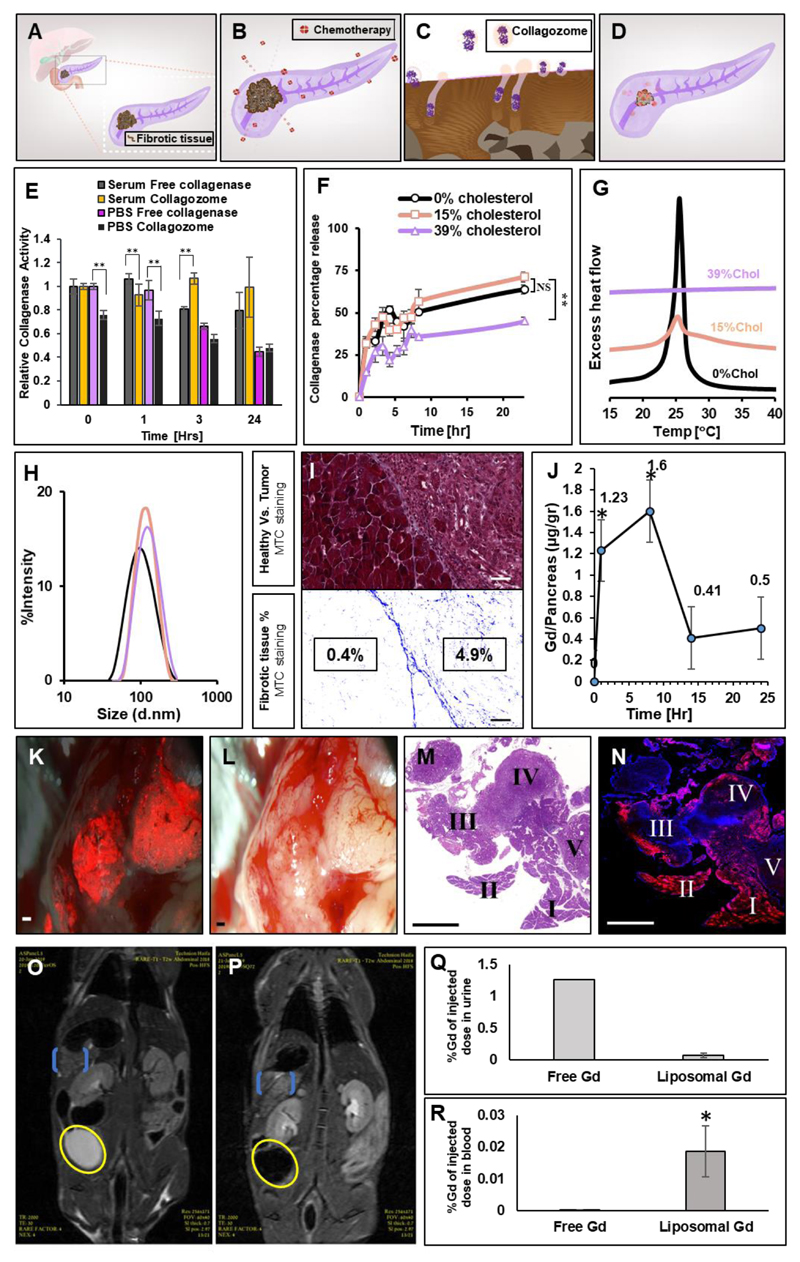
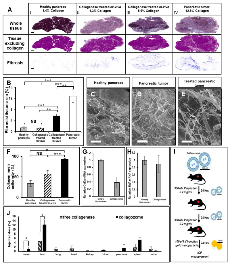
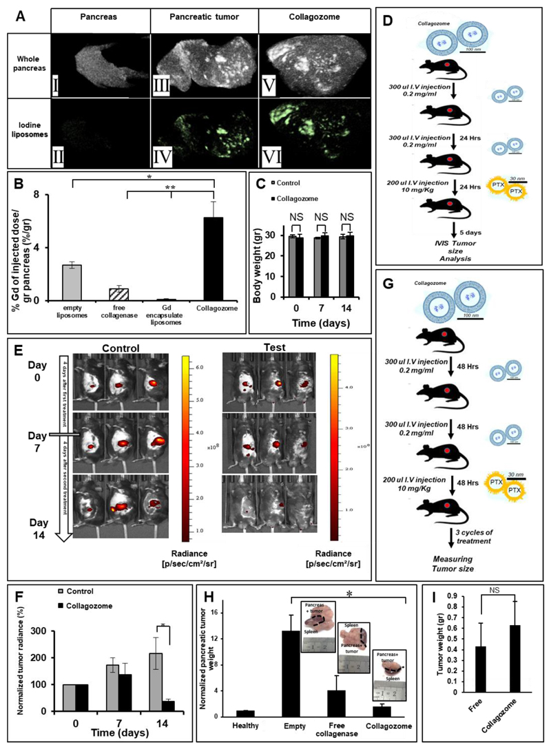
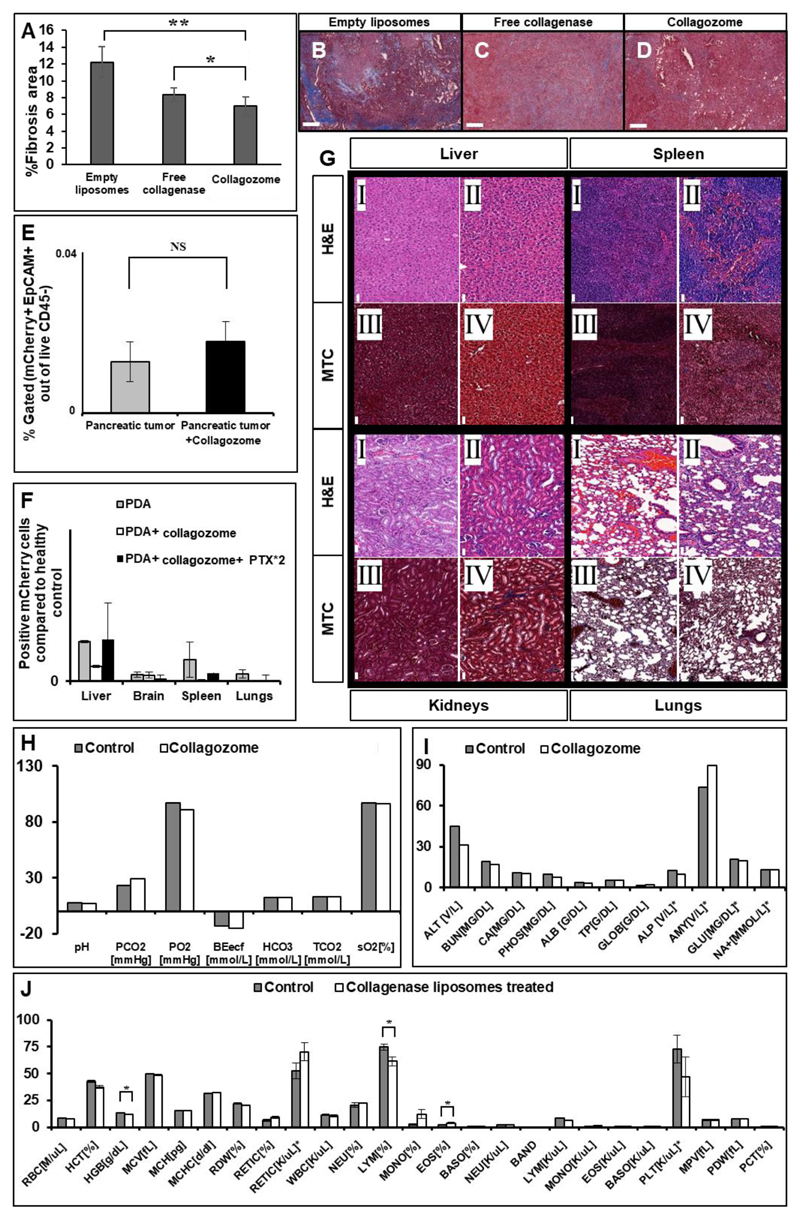
Overexpressed extracellular matrix (ECM) in pancreatic ductal adenocarcinoma (PDAC) limits drug penetration into the tumor and is associated with poor prognosis. Here, we demonstrate that a pretreatment based on a proteolytic-enzyme nanoparticle system disassembles the dense PDAC collagen stroma and increases drug penetration into the pancreatic tumor. More specifically, the collagozome, a 100 nm liposome encapsulating collagenase, was rationally designed to protect the collagenase from premature deactivation and prolonged its release rate at the target site. Collagen is the main component of the PDAC stroma, reaching 12.8 ± 2.3% vol in diseased mice pancreases, compared to 1.4 ± 0.4% in healthy mice. Upon intravenous injection of the collagozome, ∼1% of the injected dose reached the pancreas over 8 h, reducing the level of fibrotic tissue to 5.6 ± 0.8%. The collagozome pretreatment allowed increased drug penetration into the pancreas and improved PDAC treatment. PDAC tumors, pretreated with the collagozome followed by paclitaxel micelles, were 87% smaller than tumors pretreated with empty liposomes followed by paclitaxel micelles. Interestingly, degrading the ECM did not increase the number of circulating tumor cells or metastasis. This strategy holds promise for degrading the extracellular stroma in other diseases as well, such as liver fibrosis, enhancing tissue permeability before drug administration.

Keywords: collagen; extracellular matrix; fibrosis; liposome; nanoparticle; paclitaxel micelles; pancreatic cancer.


Figures

Figure 1
Figure 1. Collagenase nanoparticles and biodistribution to orthotopic pancreatic PDAC tumors.
Pancreatic ductal adenocarcinoma (PDAC) is characterized by the overexpression of extracellular matrix (ECM) – schematic outline of the study (A). The dense ECM contributes to tumor drug resistance (B). Proteolytic enzymes housed within nanoparticles were used to disassemble the collagen component of the tumor ECM (C). Collagenase encapsulated liposomes, collagozome, pretreatment increased tumor' drug uptake, allowing more effective treatment (D). Collagenase type-I encapsulation protects the enzyme from deactivation in mouse plasma. Enzyme activity was compared inside the particles and in plasma (E), (n=3) **indicates p-value between specified bars <0.01. Different liposome formulations were tested to determine the optimal collagenase nanoparticle. Liposomes were composed of DMPC: cholesterol:DSPE-PEG2000; the cholesterol content in the membrane was varied (0, 15 or 39 mole%) to test the effect on prolonging the collagenase release rate at 37°C (F), (n=3) **indicates p-value to 39% cholesterol<0.01. Differential scanning calorimetry (DSC) thermograms (G) and dynamic light scattering (DLS) size measurements (H) of DMPC liposomes containing 0, 15 and 39 mole% cholesterol, suspended in PBS pH 7.4. While a sharp transition temperature can be seen 25.5°C for the 0 mole% cholesterol formulation, no transition temperature is noticed in the 39 mole% formulation. Insignificant size differences were observed between the three liposome formulations. PDAC fibrotic collagen matrix acts as a mass transport barrier. The tumor’s ECM tissue has an increased collagen component (I). Collagen was stained with Masson’s Trichrome in healthy and diseased pancreatic tissue. Scale bar= 50μm. Poor nanoparticle pancreatic uptake was observed over 24 hours in PDAC-bearing mice (J). The amount of Gd-liposomes that reached the pancreas of PDAC mice at different time intervals after an intravenous administration was quantified using elemental analysis (n=3) *indicates p-value to control <0.05. The majority of Gd-liposomes reach the healthy region in the diseased pancreas. Fluorescent liposomes were imaged in the pancreas using intra-vital microscopy (fluorescent – K,bright field – L; scale bar= 1 mm, fluorescent liposomes are labeled red). Histological H&E-staining analysis of the pancreas enabled differentiating between the healthy (MI,II,NI,II) and diseased segments of the pancreas (MIII-V, NIII-V). Fluorescent histology of the corresponding sections (K) indicated that most of the nanoparticles are concentrated in the healthy regions of the diseased pancreas (L; Scale bar=2mm; fluorescent sections: liposomes-red, nuclei-blue, n=3). Majority of the nanoparticles are secreted within 25 minutes after intravenous injection. Gd liposomes were injected to the tail vein and the mice were flash-scanned using 9.4Tesla MRI. The Gd signal was imaged in the bladder and pancreas following 25 minutes (O) and 24 hours (P) tumor marked in blue, bladder marked in yellow. Gd quantity was measured via elemental analysis (ICP-OES) in urine (Q) and blood (R) of at least n=3 mice following 30 minutes from intravenous injection of either free Gd or Liposomal Gd. All p-values presented were calculated according to a student's t-test with a two-tailed distribution with unequal variance. 1A-D Illustrated by Dima Zagorski.
Figure 2
Figure 2. Collagenase reduces the level of fibrosis in pancreatic tumors.
Healthy pancreases (AI), collagenase treated PDAC ex-vivo (AII), collagenase-treated PDAC in-vivo (AIII) and non-treated PDAC (AIV) collagen was stained and collagen density was evaluated (B). n=8 in each treatment group. Collagen fibers in healthy, PDAC and collagenase-treated PDAC tumors in-vivo (C-E). PDAC-bearing mice and healthy mice were treated with collagozome; the pancreas was then decellularized and imaged using HR-SEM. PDAC mice after being treated with collagozome (C) and healthy pancreas (D) were compared to non-treated PDAC pancreas (E). The collagen mesh size was measured in healthy and PDAC mice, before and after the collagenase treatment. The in-vivo treatment decreased the mesh size to levels found in healthy pancreas (F). Collagen type 3 expression is reduced following collagozome pretreatment (G-H). mRNA was extracted from PDAC tumors following pretreatment with either empty or collagozome. cDNA was produced and qRTPCR was performed to determine relative expression of Collagen 3 alpha- col3a, (G) and smooth muscle actin- SMA (H). mRNA expression levels were calculated relative to GAPDH. Biodistribution of Gold nanoparticles to various organs was enhanced in healthy and tumor-baring mice following collagenase treatment (I,J) A schematic representation of the treatment protocol (I). Mice were administered intravenously with 100-nm collagozome, day after day, followed by a single dose of 100-nm gold nanoparticles (100μl, 5mg/ml) (n=5 biological replicates for each pretreatment to mice).The amount of Gold nanoparticles that reached the various organs of PDAC mice after an intravenous administration was quantified using elemental analysis. Results in tumor-baring mice injected with 100-nm gold nanoparticles (J). *indicates p-value<0.05 according to a student's t-test with two-tailed distribution with unequal variance. **indicates p-value <0.01. ***indicates p-value<0.001 according to student's t-test with a two-tailed distribution with unequal variance. All P-values presented were calculated between specified bars according to a student's t-test with a two-tailed distribution with heterodetic unequal variance for multiple comparisons.
Figure 3
Figure 3. Enhanced pancreatic uptake after the collagozome pretreatment.
Collagozome pretreatment increased the pancreatic uptake of 100-nm liposomes. Iodine-loaded (A) or Gd-loaded (B) 100-nm liposomes were injected intravenously to mice bearing PDAC tumors, and the pancreases were imaged by CT; PDAC collagozome treatment group (AV-VI) was compared to the non-treated PDAC group (AIII-IV) and to a control group (AI-II). Similar quantitative results were recoded using Gd-liposomes and elemental analysis of the pancreas. (n=10 for the empty liposome group, n=5 of the free enzyme group, n=3 for the no pretreatment group and n=4 for the Collagozome treatment group). Mice were administered 100-nm collagozome day after day, followed by a single dose of 30-nm paclitaxel-loaded micelles (10 mg/kg-body-weight) and the mCherry-expressing tumors were imaged. No major body-weight fluctuations were measured among the groups (C). Therapeutic efficacy was recorded in mice pre-treated with collagozome followed by paclitaxel (D, E, F). Mice bearing PDAC tumors pretreated (right) or non-treated (left) with collagozome, 5 days after pretreatment were treated with paclitaxel micelles (D). Tumors were imaged over two weeks (E); tumor radiance decreased by 64% in the liposome (test) group and increased by 16% in the control (F). Results are representative of five biological replicates for both groups. A schematic representation of the treatment protocol (G) Mice baring PDAC tumors were administered with 100-nm Collagozome, empty or free collagenase, in 48 hour gaps, followed by a single dose of 30-nm paclitaxel-loaded micelles (10 mg/kg-body-weight). This was repeated for 3 cycles with at least 3 mice in each experimental group. Therapeutic efficacy was recorded in mice pre-treated with collagozome liposomes followed by paclitaxel (H). Mice bearing PDAC tumors were pretreated with either collagozome, empty liposomes or free liposomes followed by paclitaxel micelle treatment. Tumors were weighed at the end point. Values presented are relative to healthy pancreas size. (H). No change in tumor weight was observed between collagozome treated mice (without paclitaxel micelle treatment) compared to free collagozome (I). *indicates p-value<0.05, **indicates p-value<0. All P-values presented were calculated between specified bars according to a student's t-test with a two-tailed distribution with heterodetic unequal variance for multiple comparisons.
Figure 4
Figure 4. Collagozome reduce collagen quantity in PDAC tumors (A-D)
Mice baring PDAC tumors were administered with either 100-nm empty liposomes (B), free collagenase (C), or collagozome (D), twice a week for three weeks. Pancreatic tumor sections were stained for collagen with Masson’s Trichrome. Scale bar= 200μm. Quantification of Masson’s Trichrome staining was done by Image J. Five sections were quantified for % collagen in tumor area in at least three mice of the same treatment (A). Circulating tumor cells (CTCs) and tissue morphology after collagenase treatments histological evaluation of organs from in-vivo collagenase-treated and untreated mice (E). H&E staining (I,II) Masson's Trichrome collagen staining (III,IV) of the liver, spleen, kidneys and lungs among the collagozome non-treated (odd numbers) and treated groups (even numbers). Results are representative of two biological replicates for both groups). Scale bar= 50μm. Circulating tumor cells (CTCs) were measured in mice serum 24-hours after the collagenase treatment (F). No significant differences were observed between the PDAC-treated and the non-treated groups (n=4 for both groups). NS indicates p-value>0.05 according to a Student's t-test with a two-tailed distribution with equal variance. No evidence of metastatic disease was observed in the liver, brain, spleen and lungs 7 days after the collagenase treatment (G). Results are representative of 2 biological replicates for all groups. NS indicates p-value>0.05 according to a Student's t-test with a two-tailed distribution with unequal variance. Comparing blood panels of collagozome treated and untreated mice: oxygen, chemistry and electrolyte analysis (H,I), red blood cell, reticulocyte, platelet count and white blood cell analysis (J) between the collagozome treated mice and non-treated mice 24 hours after the injection. *indicates p-value<0.05 All P-values presented were calculated between specified bars according to a student's t-test with a two-tailed distribution with heterodetic unequal variance for multiple comparisons.

Similar articles

Cited by

References

    1. Olive KP, Jacobetz MA, Davidson CJ, Gopinathan A, McIntyre D, Honess D, Madhu B, Goldgraben MA, Caldwell ME, Allard D, Frese KK, et al. Inhibition of Hedgehog Signaling Enhances Delivery of Chemotherapy in a Mouse Model of Pancreatic Cancer. Science. 2009;324:1457–1461. - PMC - PubMed
    1. Schuerle S, Soleimany AP, Yeh T, Anand GM, Haberli M, Fleming HE, Mirkhani N, Qiu F, Hauert S, Wang X, Nelson BJ, et al. Synthetic and Living Micropropellers for Convection-Enhanced Nanoparticle Transport. Sci Adv. 2019;5:eaav4803. - PMC - PubMed
    1. Neesse A, Algul H, Tuveson DA, Gress TM. Stromal Biology and Therapy in Pancreatic Cancer: A Changing Paradigm. Gut. 2015;64:1476–1484. - PubMed
    1. Molinaro R, Corbo C, Livingston M, Evangelopoulos M, Parodi A, Boada C, Agostini M, Tasciotti E. Inflammation and Cancer: In Medio Stat Nano. Curr Med Chem. 2018;25:4208–4422. - PMC - PubMed
    1. Provenzano PP, Cuevas C, Chang AE, Goel VK, Von Hoff DD, Hingorani SR. Enzymatic Targeting of the Stroma Ablates Physical Barriers to Treatment of Pancreatic Ductal Adenocarcinoma. Cancer Cell. 2012;21:418–429. - PMC - PubMed

Publication types

MeSH terms