Self-Activated Electrical Stimulation for Effective Hair Regeneration via a Wearable Omnidirectional Pulse Generator
- PMID: 31503449
- PMCID: PMC6881522
- DOI: 10.1021/acsnano.9b03912
Self-Activated Electrical Stimulation for Effective Hair Regeneration via a Wearable Omnidirectional Pulse Generator
Abstract
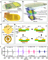
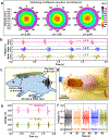
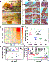
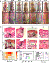
Hair loss, a common and distressing symptom, has been plaguing humans. Various pharmacological and nonpharmacological treatments have been widely studied to achieve the desired effect for hair regeneration. As a nonpharmacological physical approach, physiologically appropriate alternating electric field plays a key role in the field of regenerative tissue engineering. Here, a universal motion-activated and wearable electric stimulation device that can effectively promote hair regeneration via random body motions was designed. Significantly facilitated hair regeneration results were obtained from Sprague-Dawley rats and nude mice. Higher hair follicle density and longer hair shaft length were observed on Sprague-Dawley rats when the device was employed compared to conventional pharmacological treatments. The device can also improve the secretion of vascular endothelial growth factor and keratinocyte growth factor and thereby alleviate hair keratin disorder, increase the number of hair follicles, and promote hair regeneration on genetically defective nude mice. This work provides an effective hair regeneration strategy in the context of a nonpharmacological self-powered wearable electronic device.
Keywords: growth factors; hair regeneration; motion-activated device; physical approach; rats and nude mice.
Conflict of interest statement
The authors declare no competing financial interest.
Figures


Similar articles
-
Conductive Bio-Harvesting Tonic (CBT) with an Anti-Dandruff Effect Enhances Hair Growth by Utilizing Naturally Generated Electric Energy during Human Activities.J Microbiol Biotechnol. 2024 Nov 28;34(11):2376-2384. doi: 10.4014/jmb.2408.08014. Epub 2024 Sep 6. J Microbiol Biotechnol. 2024. PMID: 39300961 Free PMC article.
-
Hair follicle germs containing vascular endothelial cells for hair regenerative medicine.Sci Rep. 2021 Jan 12;11(1):624. doi: 10.1038/s41598-020-79722-z. Sci Rep. 2021. PMID: 33436760 Free PMC article.
-
Low-frequency electromagnetic fields promote hair follicles regeneration by injection a mixture of epidermal stem cells and dermal papilla cells.Electromagn Biol Med. 2020 Oct 1;39(4):251-256. doi: 10.1080/15368378.2020.1793165. Epub 2020 Jul 30. Electromagn Biol Med. 2020. PMID: 32727226
-
Functional complexity of hair follicle stem cell niche and therapeutic targeting of niche dysfunction for hair regeneration.J Biomed Sci. 2020 Mar 14;27(1):43. doi: 10.1186/s12929-020-0624-8. J Biomed Sci. 2020. PMID: 32171310 Free PMC article. Review.
-
The dermal papilla: an instructive niche for epithelial stem and progenitor cells in development and regeneration of the hair follicle.Cold Spring Harb Perspect Med. 2014 Jul 1;4(7):a015180. doi: 10.1101/cshperspect.a015180. Cold Spring Harb Perspect Med. 2014. PMID: 24985131 Free PMC article. Review.
Cited by
-
Wireless-Powered Electrical Bandage Contact Lens for Facilitating Corneal Wound Healing.Adv Sci (Weinh). 2022 Nov;9(31):e2202506. doi: 10.1002/advs.202202506. Epub 2022 Sep 8. Adv Sci (Weinh). 2022. PMID: 36073832 Free PMC article.
-
Electrical Stimulation Therapy - Dedicated to the Perfect Plastic Repair.Adv Sci (Weinh). 2025 Jun;12(24):e2409884. doi: 10.1002/advs.202409884. Epub 2024 Dec 16. Adv Sci (Weinh). 2025. PMID: 39680745 Free PMC article. Review.
-
Water-powered, electronics-free dressings that electrically stimulate wounds for rapid wound closure.Sci Adv. 2024 Aug 9;10(32):eado7538. doi: 10.1126/sciadv.ado7538. Epub 2024 Aug 7. Sci Adv. 2024. PMID: 39110791 Free PMC article.
-
3D-printed weight holders design and testing in mouse models of spinal cord injury.Front Drug Deliv. 2024 May 22;4:1397056. doi: 10.3389/fddev.2024.1397056. eCollection 2024. Front Drug Deliv. 2024. PMID: 40836991 Free PMC article.
-
Advanced Triboelectric Applications of Biomass-Derived Materials: A Comprehensive Review.Materials (Basel). 2024 Apr 24;17(9):1964. doi: 10.3390/ma17091964. Materials (Basel). 2024. PMID: 38730775 Free PMC article. Review.
References
-
- Lei M; Chuong CM Aging, Alopecia, and Stem cells. Science 2016, 351, 559. - PubMed
-
- Matsumura H; Mohri Y; Binh NT; Morinaga H; Fukuda M; Ito M; Kurata S; Hoeijmakers J; Nishimura EK Hair Follicle Aging is Driven by Transepidermal Elimination of Stem Cells via COL17A1 Proteolysis. Science 2016, 351, aad4395. - PubMed
-
- Price VH Treatment of Hair Loss. N. Engl. J. Med 1999, 341, 964. - PubMed
-
- Marks DH; Penzi LR; Ibler E; Manatis-Lornell A; Hagigeorges D; Yasuda M; Drake LA; Senna MM The Medical and Psychosocial Associations of Alopecia: Recognizing Hair Loss as More Than a Cosmetic Concern. Am. J. Clin. Dermatol 2019, 20, 195–200. - PubMed
-
- Sasson M; Shupack JL; Stiller MJ Status of Medical Treatment for Androgenetic Alopecia. Int. J. Dermatol 1993, 32, 701–706. - PubMed
Publication types
MeSH terms
Grants and funding
LinkOut - more resources
Full Text Sources
Research Materials

