. 2019 Sep 10;14(9):e0221550.
doi: 10.1371/journal.pone.0221550. eCollection 2019.

Identification of variant HIV envelope proteins with enhanced affinities for precursors to anti-gp41 broadly neutralizing antibodies

Affiliations

Identification of variant HIV envelope proteins with enhanced affinities for precursors to anti-gp41 broadly neutralizing antibodies

Hong Zhu et al. PLoS One. .

Abstract

HIV envelope protein (Env) is the sole target of broadly neutralizing antibodies (BNAbs) that are capable of neutralizing diverse strains of HIV. While BNAbs develop spontaneously in a subset of HIV-infected patients, efforts to design an envelope protein-based immunogen to elicit broadly neutralizing antibody responses have so far been unsuccessful. It is hypothesized that a primary barrier to eliciting BNAbs is the fact that HIV envelope proteins bind poorly to the germline-encoded unmutated common ancestor (UCA) precursors to BNAbs. To identify variant forms of Env with increased affinities for the UCA forms of BNAbs 4E10 and 10E8, which target the Membrane Proximal External Region (MPER) of Env, libraries of randomly mutated Env variants were expressed in a yeast surface display system and screened using fluorescence activated cell sorting for cells displaying variants with enhanced abilities to bind the UCA antibodies. Based on analyses of individual clones obtained from the screen and on next-generation sequencing of sorted libraries, distinct but partially overlapping sets of amino acid substitutions conferring enhanced UCA antibody binding were identified. These were particularly enriched in substitutions of arginine for highly conserved tryptophan residues. The UCA-binding variants also generally exhibited enhanced binding to the mature forms of anti-MPER antibodies. Mapping of the identified substitutions into available structures of Env suggest that they may act by destabilizing both the initial pre-fusion conformation and the six-helix bundle involved in fusion of the viral and cell membranes, as well as providing new or expanded epitopes with increased accessibility for the UCA antibodies.


Conflict of interest statement

The authors have read the journal's policy and the authors of this manuscript have the following competing interest: JZ was supported by NIH grant R01GM076485, which was awarded to Dr. David Mathews. Dr. David Mathews is Principal Investigator on R01GM076485 but is not a listed author. This does not alter the authors' adherence to PLOS ONE policies on sharing data and materials.

Figures
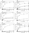
Fig 1
Fig 1
a) Saturation binding of 4E10 UCA to reconstructed QH0692dsm Env variants related to sorted clone QH-17. b) Saturation binding of 4E10 UCA to reconstructed QH0692dsm Env variants related to sorted clone P6-8. c) Saturation binding of 4E10 UCA to reconstructed QH0692dsm Env variants carrying substitutions at position W631. d) Saturation binding of 4E10 UCA to reconstructed QH0692dsm and YU2dsm Env variants carrying substitutions at position W666. e) Saturation binding of 4E10 UCA to QH0692dsm Env variants carrying substitutions at position I642. f) Saturation binding of 4E10 UCA to QH0692dsm Env variants carrying combined mutations.
Fig 2
Fig 2
a) Saturation binding assay of 10E8 UCA to reconstructed YU2dsm Env variants corresponding to clones C7, C38, and C47 from 10E8 UCA-sorted library (containing the amino acid substitutions noted on figure). b) Saturation binding assay of 10E8 UCA to reconstructed YU2dsm Env variants carrying mutations W666R and W666K to 10E8 UCA. c) Saturation binding assay of 10E8 UCA to reconstructed library clone C12 lacking W666R but containing the substitutions P498H, S534A, L566M, W614R, N637T, Y638N, Y643H, Q648R, and A667S.
Fig 3
Fig 3
a) Effect of truncation (after W670) or mutagenesis (substitutions W672A F673A T675A) of the epitope for mature 4E10 on the binding of mature 4E10 to variant and otherwise unmutagenized forms of QH0692 gp140dsm; b) Effect of truncation (after W670) or mutagenesis (substitutions W672A F673A T675A) of the epitope for mature 4E10 on binding of the 4E10 UCA to variant and otherwise unmutagenized forms of QH0692 gp140dsm. c) Effect of truncation (after W670) of the epitope for mature 10E8 on binding of the 10E8 UCA to YU2 gp140dsm either containing or lacking the W666R substitution. d) Effect of truncation (after W670) of the epitope for mature 10E8 on binding of mature 10E8 to YU2 gp140dsm either containing or lacking the W666R substitution.
Fig 4
Fig 4
a) Nine of the ten most frequently identified mutations conferring Env binding to the 4E10 UCA map to the “clasp” region of Env that is proposed to lock the protein in the pre-fusion state. a) Sites corresponding to mutations Q619R, W623R, W631R, and W628R, four of the most abundant mutations from the NGS sequencing of the sorted library (see Table 2) are located directly in the M530-tryptophan clasp. The mutated residues are indicated in color-coded stick representations corresponding to the residues indicated on the figure. Residues M530 and C605 (as a disulfide with C501) are also indicated for reference. The displayed structure is the presumed pre-fusion form of the BG505 SOSIP.664 Env Trimer (PDB code: 4TVP [56]) in complex with antibodies PGT122 and 35O22 (not shown in this figure). Note that position 619 is a serine in Env from strain BG505, as indicated, but is glutamine in Env of strain QH0692. Gp41 is shown in green, gp120 in dark purple. b) Sites corresponding to mutations C605R, W610R, W614R, Y638H, L646S, map to the interface between gp41 and the N- and C-terminal loop of gp120 near the M530 clasp (same view of Env as panel a, with different residues highlighted).
Fig 5
Fig 5. Trp-rich sequence motifs in the gp41 region of Env.
The figure was generated using the AnalyzeAlign database of the Los Alamos National Laboratory (http://www.hiv.lanl.gov/) based on the curated alignment of 6636 sequences. Trp residues are shown in red, Gln residues in green and Ile residues in blue. Abundances of selected residues are shown in S3 Table.
Fig 6
Fig 6. Frequently identified mutations conferring Env binding to the 4E10 UCA map to the hydrophobic groove between adjacent helices in the six-helix, fusion-active form of gp41.
a) Mutated residues indicated on the structure of protease cleaved gp41, which consists of a complex of peptides corresponding to residues 546–581 and 528–661 of Env (HXB2 numbering). Two helices from the overall six helix structure (PDB code: 1AIK [49]) are shown. This structure is reported to be very similar to other structures of putative fusion-active forms of gp41 [56]. The sites of the W628R, W631R, I635, Y638H, and L646S, frequently-recovered mutations among NGS reads are indicated as color-coded stick representations. Note that other frequently-recovered mutations are at sites outside the region of sequence encompassed by this structure. One additional indicated site, I642, was recovered less frequently, but was shown to modulate binding to the 4E10 UCA when mutated together with more frequently recovered substitutions (see text). b) 90° rotated view of the structure shown in a).
Fig 7
Fig 7. W666R, the most frequently recovered mutation conferring binding to the 10E8 UCA maps to the trimer interface of the putative pre-fusion structure of the MPER region and the adjacent transmembrane domain.
The figure is based on the NMR structure of residues 660–710 (HXB2 numbering) of Env from HIV strain 92UG024.2 reconstituted into dimyristoylphophatidylcholine/dihexanoylphosphatiylcholine (DMPC/DHPC) bicelles. (PDB code 6E8W [48]). a) The position of W666R is shown as a stick representation in red. The two positions (I682 and R683) of three mutations that were frequently recovered in the 10E8 UCA-sorted library are also indicated. These are the only three of the most abundant mutations (Table 5) that map to the sequences included in this structure. Note that position 683 is occupied by lysine in the envelope from the YU2 strain but is arginine in Env from strain 92UG024.2. a) Representation viewed parallel to the membrane surface. b) Representation viewed perpendicular to the membrane.

Similar articles

References

    1. Fauci AS. An HIV Vaccine Is Essential for Ending the HIV/AIDS Pandemic. JAMA. 2017;318(16):1535–6. 10.1001/jama.2017.13505 - DOI - PubMed
    1. Gao Y, McKay PF, Mann JFS. Advances in HIV-1 Vaccine Development. Viruses. 2018;10(4). - PMC - PubMed
    1. Rerks-Ngarm S, Pitisuttithum P, Nitayaphan S, Kaewkungwal J, Chiu J, Paris R, et al. Vaccination with ALVAC and AIDSVAX to prevent HIV-1 infection in Thailand. N Engl J Med. 2009;361(23):2209–20. 10.1056/NEJMoa0908492 - DOI - PubMed
    1. Kwong PD, Mascola JR. HIV-1 Vaccines Based on Antibody Identification, B Cell Ontogeny, and Epitope Structure. Immunity. 2018;48(5):855–71. 10.1016/j.immuni.2018.04.029 - DOI - PubMed
    1. Huang J, Ofek G, Laub L, Louder MK, Doria-Rose NA, Longo NS, et al. Broad and potent neutralization of HIV-1 by a gp41-specific human antibody. Nature. 2012;491(7424):406–12. 10.1038/nature11544 - DOI - PMC - PubMed

Publication types

MeSH terms

LinkOut - more resources