. 2019 Sep 10;15(9):e1007936.
doi: 10.1371/journal.ppat.1007936. eCollection 2019 Sep.

The phage gene wmk is a candidate for male killing by a bacterial endosymbiont

Affiliations

The phage gene wmk is a candidate for male killing by a bacterial endosymbiont

Jessamyn I Perlmutter et al. PLoS Pathog. .

Abstract

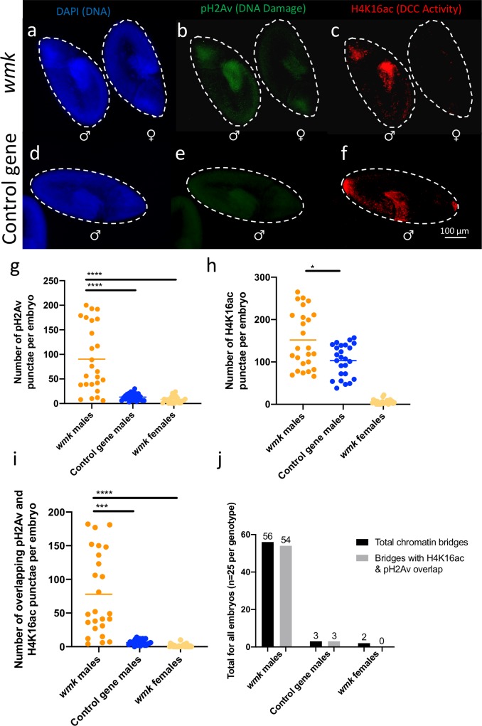
Wolbachia are the most widespread maternally-transmitted bacteria in the animal kingdom. Their global spread in arthropods and varied impacts on animal physiology, evolution, and vector control are in part due to parasitic drive systems that enhance the fitness of infected females, the transmitting sex of Wolbachia. Male killing is one common drive mechanism wherein the sons of infected females are selectively killed. Despite decades of research, the gene(s) underlying Wolbachia-induced male killing remain unknown. Here using comparative genomic, transgenic, and cytological approaches in fruit flies, we identify a candidate gene in the eukaryotic association module of Wolbachia prophage WO, termed WO-mediated killing (wmk), which transgenically causes male-specific lethality during early embryogenesis and cytological defects typical of the pathology of male killing. The discovery of wmk establishes new hypotheses for the potential role of phage genes in sex-specific lethality, including the control of arthropod pests and vectors.


Conflict of interest statement

J.I.P. and Seth R.B. are listed as inventors on a patent related to potential applications of wmk in arthropods.

Figures

Fig 1
Fig 1. Comparative genomics of wmk and its homologs in wMel and male-killing strains.
Prophage WO gene regions containing wmk, wmk-like homologs, and CI genes cifA and cifB are listed by Wolbachia strain in bold and then prophage. At least one wmk homolog is associated with each Wolbachia-induced male killing strain. Genes pointing in the same direction are on the same DNA strand. The distance between wmk and cifA is approximately 5 kb. Shading highlights homologs in each strain. (*) wmk homologs are annotated as transcriptional regulators in the Wolbachia reference genomes and encode helix-turn-helix XRE domains (S4 Table). (**) While wBif reportedly induces weak CI after temperature treatment [8], the assembled genome does not contain cifB.
Fig 2
Fig 2. Transgenic expression of wmk causes a female-biased sex ratio.
Each sample point represents the adult offspring produced by a replicate family of ten mothers and two fathers (average offspring number per data point is 90). Bars represent the average sex ratio. Control gene flies have the Wolbachia transgene WD0034. WT is the BSC8622 strain. E = expressing, NE = non-expressing, Act5c has an Act5c-Gal4 gene, CyO has the CyO chromosome. wmk-expressing flies have a significantly female-biased sex ratio against all other genotypes. This experiment has been done four times. Statistics are based on a Kruskal-Wallis one-way ANOVA followed by Dunn’s correction. **p<0.01, *** p<0.001. Orange dots represent wmk, blue dots represent the control gene, and gray dots represent the WT strain.
Fig 3
Fig 3. Transgenic expression of wmk causes cytological defects in early embryogenesis.
Data are from pooled embryos (both sexes, expressing and non-expressing) with either wmk, the control gene, or an uninfected wild type (WT) background (see methods). (A-C) Defective wmk embryos fixed 3–4 h after egg deposition (AED) exhibit either chromatin bridging (arrowheads), pyknotic nuclei, or local mitotic failure leading to gaps in the distribution of nuclei, respectively. (B) Image has been brightened for visibility. (D) Image of a normal control gene embryo fixed 3–4 h AED. (E) Image of unfertilized embryo fixed approximately 3–4 h AED. (F) Image of degraded wmk embryo fixed 16–17 h AED with no distinct nuclei and no visible segmentation. (G) Image of a degraded wmk embryo fixed 16–17 h AED with no distinct nuclei, but the cephalic furrow is (indicated by arrowheads). (F) and (G) are brightened in order to see their differences. (H) Image of normal control gene embryo fixed 16–17 h AED. (I) Graph quantitating the percentage of embryos exhibiting DNA defects that were fixed 1–2 h AED. N = 220 for the wmk cross, N = 200 for the control gene cross, and N = 169 for the WT cross. Total refers to the total percentage of embryos with one or more of the three defects (embryos can have more than one, as in (A)). All differences within each defect category were not statistically significant. (J) Graph of the percentage of embryos exhibiting DNA defects that were fixed 3–4 h AED for wmk, control gene, and WT crosses. N = 276 for the wmk cross, N = 273 for the WT cross, and N = 279 for the control transgene cross. (K) Graph of the percentage of degraded embryos fixed 16–17 h AED in the wmk, control gene, and WT crosses. N = 327 for the wmk cross, N = 315 for the control transgene cross, and N = 231 for the WT cross. The percent of unfertilized eggs is the expected percent given the observed rate of unfertilized sibling eggs fixed 3–4 h AED (wmk, 8%, N = 324; control gene, 4.5%, N = 202; WT, 7%, N = 217). Statistics for (I), (J), and (K) were performed with a Chi-square test comparing the three genotypes within each defect category. These experiments have been performed once. The white border around (F, G, & H) indicates embryos fixed 16–17 h AED, while the rest (A-E) are embryos fixed 3–4 h AED. All images were taken at 20X zoom, except the inset image in (A) that is a zoomed in image of the same region. ** p<0.01, *** p<0.001, ****p<0.0001.
Fig 4
Fig 4. wmk-induced embryonic defects are enriched in males.
Data are from pooled embryos (both sexes, expressing and non-expressing, see methods) with either wmk, the control gene, or a WT background. (A) Graph quantitating the percentage of 3–4 h AED embryos (males or females) that have at least one defect (wmk males N = 228, control gene males N = 190, WT males N = 170, wmk females N = 240, control gene females N = 200, WT females N = 158). (B) Graph quantitating the sex ratio of viable embryos (not degraded, no visible defects) across two development times (1–2 h wmk, N = 105 m, 111 f; 1–2 h control gene, N = 30 m, 141 f; 1–2 h WT, N = 112 m, 115 f; 16–17 h wmk, N = 104 m, 154 f; 16–17 h control gene, N = 116 m, 120 f; 16017 h WT, N = 110 m, 108 f). m = male, f = female. Statistics were performed with a Chi-square test comparing the three genotypes within each category (male or female in (A) and 1–2 h or 16–17 h in (B)). These experiments were performed once. ** p<0.01, *** p<0.001, ****p<0.0001.
Fig 5
Fig 5. Transgenic expression of wmk causes DNA damage in association with H4K16ac.
Images and data are from embryos 4–5 h AED expressing a transgene under the arm driver. (A) DAPI DNA stain of male and female embryos, side-by-side, expressing wmk. Sexes determined by H4K16ac antibody. (B) pH2Av antibody staining of the same embryos as (A). The male has distinct punctae or foci, while the female does not. All embryos exhibit either a low level of autofluorescence at the same wavelength as the secondary antibody (Alexa 488) visible in both embryos or there is background staining. (C) H4K16ac antibody staining of the same embryos as (A). Distinct punctae are only visible in males, while females can exhibit low levels of staining. (D) DAPI DNA stain of control gene male. Sex determined by H4K16ac antibody. (E) pH2Av antibody staining of the same embryo as (D), with no distinct punctae and only autofluorescence or background staining visible. (F) H4K16ac antibody staining of the same embryos as (D). (G) Graph of the number of pH2Av punctae visible in each embryo. N = 25 embryos per genotype. Statistics are based on a Kruskal-Wallis one-way ANOVA followed by Dunn’s correction. (H) Graph of the number of H4K16ac punctae visible in each of the same embryos as measured in (G). Statistics are based on a Mann-Whitney U test comparing the two male categories. (I) Number of cases where pH2Av punctae directly overlapped with H4K16ac punctae in the same embryos as (G) and (H). Statistics are based on a Kruskal-Wallis one-way ANOVA followed by Dunn’s correction. (J) Graph of the total number of chromatin bridges and the total number of bridges with overlapping H4K16ac and pH2Av punctae in each of the three genotypes measured in (G-I). All images were taken at 20X zoom. This experiment has been performed once. *p<0.05, ***p<0.001, ****p<0.0001.
Fig 6
Fig 6. Native Wolbachia gene and transgene expression in embryos of D. melanogaster and D. bifasciata.
(A) Graph of native prophage WO and Wolbachia gene expression in wMel-infected D. melanogaster embryos fixed 4–5 h AED (pooled male & female) compared to Wolbachia groEL. Each point (n = 7) represents a pool of 30 embryos from a set of 10 mothers and 2 fathers. (B) Graph of (i) transgene expression in uninfected D. melanogaster embryos fixed 4–5 h AED versus (ii) native gene expression in samples from a, both compared to Drosophila rpl36 (pooled male, female, expressing, and non-expressing for transgenes). Each point (transgene n = 8, native n = 7) represents a pool of 30 embryos from a set of 10 mothers and 2 fathers. (C) Graph of wBif Wolbachia gene expression in D. bifasciata embryos 4–5 h AED (pooled male & female) compared to Wolbachia groEL. Homologs to the control gene in this study and cifB were not measured as they are not present in the wBif genome assembly. Each point (n = 7) represents a pool of 30 embryos from a set of 10 mothers and 2 fathers. Values denote 2-ΔCt. Statistics are based on a Kruskal-Wallis one-way ANOVA followed by Dunn’s correction. This experiment has been done once. **p<0.01, ***p<0.001.

References

    1. Zug R, Hammerstein P. Still a host of hosts for Wolbachia: analysis of recent data suggests that 40% of terrestrial arthropod species are infected. PLoS One. 2012;7(6):e38544 Epub 2012/06/12. 10.1371/journal.pone.0038544 - DOI - PMC - PubMed
    1. Weinert LA, Araujo-Jnr EV, Ahmed MZ, Welch JJ. The incidence of bacterial endosymbionts in terrestrial arthropods. Proceedings of the Royal Society B: Biological Sciences. 2015;282(1807):20150249 10.1098/rspb.2015.0249 - DOI - PMC - PubMed
    1. Ferri E, Bain O, Barbuto M, Martin C, Lo N, Uni S, et al. New insights into the evolution of Wolbachia infections in filarial nematodes inferred from a large range of screened species. PLoS One. 2011;6(6):e20843 Epub 2011/07/07. 10.1371/journal.pone.0020843 - DOI - PMC - PubMed
    1. Funkhouser-Jones LJ, van Opstal EJ, Sharma A, Bordenstein SR. The Maternal Effect Gene Wds Controls Wolbachia Titer in Nasonia. Curr Biol. 2018;28(11):1692–702 e6. Epub 2018/05/22. 10.1016/j.cub.2018.04.010 - DOI - PMC - PubMed
    1. Ferree PM, Frydman HM, Li JM, Cao J, Wieschaus E, Sullivan W. Wolbachia utilizes host microtubules and Dynein for anterior localization in the Drosophila oocyte. PLoS pathogens. 2005;1(2):e14 Epub 2005/10/18. 10.1371/journal.ppat.0010014 - DOI - PMC - PubMed

Publication types

MeSH terms

LinkOut - more resources