. 2019 Oct 1;142(10):3243-3264.
doi: 10.1093/brain/awz241.

CSF1R inhibitor JNJ-40346527 attenuates microglial proliferation and neurodegeneration in P301S mice

Collaborators, Affiliations

CSF1R inhibitor JNJ-40346527 attenuates microglial proliferation and neurodegeneration in P301S mice

Renzo Mancuso et al. Brain. .

Abstract

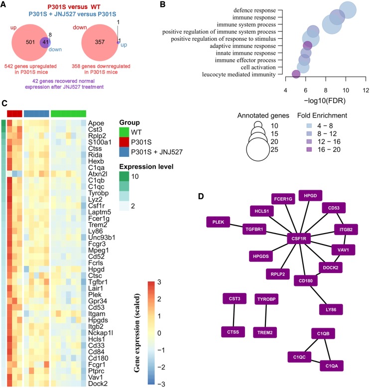
Neuroinflammation and microglial activation are significant processes in Alzheimer's disease pathology. Recent genome-wide association studies have highlighted multiple immune-related genes in association with Alzheimer's disease, and experimental data have demonstrated microglial proliferation as a significant component of the neuropathology. In this study, we tested the efficacy of the selective CSF1R inhibitor JNJ-40346527 (JNJ-527) in the P301S mouse tauopathy model. We first demonstrated the anti-proliferative effects of JNJ-527 on microglia in the ME7 prion model, and its impact on the inflammatory profile, and provided potential CNS biomarkers for clinical investigation with the compound, including pharmacokinetic/pharmacodynamics and efficacy assessment by TSPO autoradiography and CSF proteomics. Then, we showed for the first time that blockade of microglial proliferation and modification of microglial phenotype leads to an attenuation of tau-induced neurodegeneration and results in functional improvement in P301S mice. Overall, this work strongly supports the potential for inhibition of CSF1R as a target for the treatment of Alzheimer's disease and other tau-mediated neurodegenerative diseases.

Keywords: Alzheimer’s disease; CSF1R; microglia; neuroinflammation; tau.


Figures

Figure 1
Figure 1
JNJ-527 prevents CSF1R phosphorylation in vitro and blocks microglial proliferation in vivo. (A) Representative western blot of N13 cells pretreated with increasing concentrations of JNJ-527 and stimulated with CSF1. (B) Quantification shows that JNJ-527 inhibits CSF1-R phosphorylation and downstream ERK1/2 activation at concentrations higher than 10 nM. Experiments were performed in triplicate, and values were corrected by the untreated control (white bars) and expressed as mean ± SEM. (C) JNJ-527 IC50 is within 18.6 and 22.5 nM. (D) Schematic representation of the experimental design used for assessing JNJ-527 efficacy in blocking microglial proliferation (NBH n = 8, NBH+JNJ-527 30 mg/kg n = 6, ME7 n = 14, ME7+JNJ-527 3 mg/kg n = 6, ME7+JNJ-527 10 mg/kg n = 6, ME7+JNJ-527 30 mg/kg n = 8, ME7+JNJ-527 100 mg/kg n = 6). Mice were treated with JNJ-527 at different doses (3, 10, 30 and 100 mg/kg) for 5 days, with four daily injections of BrdU to quantify microglial proliferation. (E) Levels of the JNJ-527 were analysed at the end of the treatment. Both plasma and brain concentration linearly correlated with the treatment dose, with T/P ratios of 0.5–1 (see numbers above columns). (F and G) JNJ-527 treatment significantly inhibited microglial proliferation in ME7 mice. Images represent Iba-1 (DAB, brown) and BrdU (alkaline phosphatese, blue) immunohistochemistry with arrows pointing to Iba-1+ BrdU+ proliferating microglia. Quantification of proliferating microglia (Iba-1+ BrdU+ cells/mm2) showed a significant reduction at all doses tested. Subsequent analysis between different doses revealed a grester effect at 30 and 100 mg/kg. (H) Pharmacokinetic/pharmacodynamics (PK/PD) model generated from the microglial proliferation data showed an EC50 of 196/ml and 69 ng/g calculated from plasmatic and brain compound concentration, respectively. (*P < 0.05, **P < 0.01 and ***P < 0.001 versus NBH; #P < 0.05, ##P < 0.01 and ###P < 0.001 versus ME7).
Figure 2
Figure 2
Inhibition of CSF1R by JNJ-527 reduces microglial expansion and restores behavioural alterations in ME7 mice. (A) Long-term JNJ-527 resulted in a significant reduction in the number of microglia (PU.1) in CA1 of the hippocampus. Representative images of PU.1 staining in CA1 hippocampus of NBH, ME7 and ME7+JNJ-527 mice. Scale bar = 50 μm. (B) JNJ-527 treatment diminished the expression of IL1-β and (C) attenuated the hyperactive behaviour (locomotor activity) and completely prevented the onset of motor deficits (inverted screen and horizontal bar). For all quantifications, NBH n = 10, ME7 n = 8, ME7+JNJ-527 n = 9. Values are mean ± SEM. *P < 0.05 ***P < 0.001 versus NBH; #P < 0.05 versus ME7.
Figure 3
Figure 3
Target engagement and efficacy readouts for clinical testing of JNJ-527. (A and B) Quantification of CSF1 in plasma was used as a measure of peripheral target engagement. (A) Short-term (5 days) administration of JNJ-527 administration induced an increase of plasma CSF1 (NBH n = 8, ME7+JNJ-527 10 mg/kg n = 6, ME7+JNJ-527 30 mg/kg). (B) Long-term JNJ-527 administration (4 weeks) showed similar increments in plasma CSF1, reflecting consistent and long lasting target engagement in the systemic compartment (NBH n = 10, ME7 n = 8, ME7+JNJ-527 n = 9). (C) Assessment of target engagement by JNJ-527 in the CNS. CSF high dimensionality proteomics with aptamer capture arrays showed a dose dependent alteration in IL-34 and SEMA3E (FDR P < 9.19 × 10⁻¹³), among 67 other proteins (NBH n = 29, ME7 n = 30, with each type receiving JNJ-527 doses: vehicle n = 10; 10 mg/kg n = 10; 30 mg/kg n = 9, 10). (D) Analysis of long-term efficacy by TSPO imaging after long-term treatment with JNJ-527. TSPO autoradiography with [3H]-PK11195 performed in different brain regions showed a consistent increase of signal in ME7 versus NBH mice, and an attenuation after treatment with JNJ-527 (NBH n = 10, ME7 n = 8, ME7+JNJ-527 n = 9). For all graphs, values are mean ± SEM. *P < 0.05, **P < 0.01, ***P < 0.001 versus NBH; #P < 0.05 versus ME7.
Figure 4
Figure 4
Tau phosphorylation and degeneration of motor neurons is accompanied by an inflammatory reaction in the spinal cord of P301S mice. (A) Significant increase in the number of microglia in the spinal cord of 20-week-old P301S mice, with no evidence of (B) infiltration of monocytes or (C) T cells [CD11b+ CD45+ cells, wild-type (WT)-20 weeks n = 13, P301S-6 weeks n = 6, P301S-12 weeks n = 8, P301S-20 weeks n = 6]. (D) The increase in the number of microglia is similar between different regions of the spinal cord. Scale bar = 100 μm. Higher magnification in boxes are shown in 14. Scale bar = 25 μm (WT-20 weeks n = 5, P301S-6weeks n = 4, P301S-12weeks n = 5, P301S-20 weeks n = 4). (E) Morphological changes in microglia precede the increase in their number both in the white and grey matter of P301S spinal cord. Scale bar = 200 μm. Higher magnification in boxes are shown in 1–4. Scale bar, 25 μm (wild-type-20 weeks n = 5, P301S-6weeks n = 4, P301S-12weeks n = 5, P301S-20 weeks n = 4). (F) IL-1β and TNFα are significantly increased in the spinal cord of P301S mice, correlating with the degeneration of motor neurons (wild-type-20 weeks n = 10, P301S-6 weeks n = 5, P301S-12 weeks n = 4, P301S-20 weeks n = 4). (G and H) Activation of JNK (phospho-JNK) in the spinal cord of wild-type and P301S mice (wild-type-20 weeks n = 5, P301S-6 weeks n = 4, P301S-12 weeks n = 5, P301S-20 weeks n = 4). (G) Cytoplasmic accumulation of phospho-JNK in motor neurons starts by 12 weeks of age (arrows) and later affects both motor neurons and other smaller populations of neurons in the spinal cord. (H) Activation of p38 (phospho-p38) shows a similar pattern, with a translocation from the nucleus (asterisk) to the cytoplasm (arrows) of motor neurons of P301S mice from 12 weeks of age. Scale bar = 20 μm. Values are mean ± SEM. *P < 0.05, **P < 0.01 and P < 0.001 versus wild-type.
Figure 5
Figure 5
JNJ-527 reduces microglial expansion and the expression of pro-inflammatory cytokines in P301S mice. (A) Schematic representation of the experimental design used to test the efficacy of CSF1-R blockade in P301S mice. Animals were treated for 8 weeks with JNJ-527 at a dose of 30 mg/kg. (B) JNJ-527 treatment reduced the number of CD11b+ CD45+ cells in the spinal cord of P301S mice [wild-type (WT) n = 7, P301S n = 11, P301S+JNJ-527 n = 6]. (C) JNJ-527 treated mice showed a significant reduction in the expression of genes associated with microglial proliferation (wild-type n = 10, P301S n = 8, P301S+JNJ-527 n = 9). (D and E) The reduction in the number of microglia was correlated with a reduction of IL1-β and TNFα at (D) RNA and (E) protein level (wild-type n = 10, P301S n = 8, P301S+JNJ-527 n = 9). (F and G) Diminished expression of inflammatory cytokines correlated with reduced activation of JNK and p38 in the cytoplasm of spinal motor neurons of 16 weeks old P301S mice (P301S n = 9, P301S+JNJ-527 n = 10). Scale bar = 50 μm. Values are mean ± SEM. *P < 0.05, **P < 0.01 and ***P < 0.001 versus P301S.
Figure 6
Figure 6
Chronic blockade of CSF1R impacts tau pathology, prevents spinal motor neuron degeneration and improves motor function of P301S mice. JNJ-527 administration reduced (A) tau phosphorylation (AT8) and (B) aggregation in the form of neurofibrillary tangles (neurofibrillary tangles, P301S n = 4, P301S+JNJ-527 n = 5) and globular tau oligomers (globular tau oligomers, P301S n = 5, P301S+JNJ-527 n = 5). Values are mean ± SEM. *P < 0.05, **P < 0.01 and ***P < 0.001 versus P301S. (C) JNJ-527 treatment induced a trend of tau aggregation reduction as measured by MSD and did not show an effect on total tau. Graphs are represented as mean ± SEM and the P-value from an unpaired t-test is indicated in the respective graphs, with near significant values outlined (P301S n = 9, P301S+JNJ-527 n = 10). (D) Quantification of motor neuron by Nissl staining showed increased neuronal preservation in L4–L5 spinal segments of 16-weeks-old P301S mice after JNJ-527 treatment (wild-type n = 10, P301S n = 9, P301S+JNJ-527 n = 10), whereas (E) rotarod test showed improved motor function in JNJ-527 treated mice (wild-type n = 20, P301S n = 20, P301S+JNJ-527 n = 21). Scale bar = 50 μm. For the neuronal survival and functional analysis, values are mean ± SEM. ***P < 0.001 versus wild type; ##P < 0.01 and ###P < 0.001 JNJ-527 versus P301S.
Figure 7
Figure 7
Differentially expressed genes driven by JNJ-527 in the spinal cord of P301S mice. (A) Venn diagrams showing opposing patterns of expression in disease (P301S versus wild-type) and treatment (P301S+JNJ-527 versus P301S) in the spinal cord (wild-type n = 7, P301S n = 3, P301S+JNJ-527 n = 5). Briefly, 542 genes were upregulated (left) and 358 were downregulated (right) in the spinal cord of P301S mice. Of those genes, 42 recovered normal expression levels after chronic treatment with JNJ-527 (in purple). (B) FDR for top 10 biological processes overrepresented among the 41 genes upregulated in disease and downregulated with JNJ-527 treatment, the size of the circles reflects the number of genes annotated in each gene ontology (GO) term while the colour indicates the fold enrichment. (C) Heat map showing the gene expression patterns in the spinal cord samples for the overlapping genes shown in A, genes are ordered from top to bottom according to their average expression [log2 (TPM + 1)]. (D) From the aforementioned 41 genes, we selected those with a one-to-one orthologue in human and looked for known PPIs among them. The network shows the 22 known PPIs among the targeted genes, with CSF1R as a relevant hub.

References

    1. Akiyama H, Barger S, Barnum S, Bradt B, Bauer J, Cole GM et al. . Inflammation and Alzheimer’s disease. Neurobiol Aging 2000; 21: 383–421. - PMC - PubMed
    1. Akiyama H, Nishimura T, Kondo H, Ikeda K, Hayashi Y, McGeer PL. Expression of the receptor for macrophage colony stimulating factor by brain microglia and its upregulation in brains of patients with Alzheimer’s disease and amyotrophic lateral sclerosis. Brain Res 1994; 639: 171–4. - PubMed
    1. Allen B, Ingram E, Takao M, Smith MJ, Jakes R, Virdee K et al. . Abundant tau filaments and nonapoptotic neurodegeneration in transgenic mice expressing human P301S tau protein. J Neurosci 2002; 22: 9340–51. - PMC - PubMed
    1. Asai H, Ikezu S, Tsunoda S, Medalla M, Luebke J, Haydar T et al. . Depletion of microglia and inhibition of exosome synthesis halt tau propagation. Nat Neurosci 2015; 18: 1584–93. - PMC - PubMed
    1. Bellucci A, Westwood AJ, Ingram E, Casamenti F, Goedert M, Spillantini MG. Induction of inflammatory mediators and microglial activation in mice transgenic for mutant human P301S tau protein. Am J Pathol 2004; 165: 1643–52. - PMC - PubMed

Publication types

MeSH terms