. 2020 Jul 1;116(8):1525-1538.
doi: 10.1093/cvr/cvz238.

Endothelial microvesicles carrying Src-rich cargo impair adherens junction integrity and cytoskeleton homeostasis

Affiliations

Endothelial microvesicles carrying Src-rich cargo impair adherens junction integrity and cytoskeleton homeostasis

Victor Chatterjee et al. Cardiovasc Res. .

Abstract

Aims: Microvesicles (MVs) conduct intercellular communication and impact diverse biological processes by transferring bioactive cargos to other cells. We investigated whether and how endothelial production of MVs contribute to vascular dysfunction during inflammation.

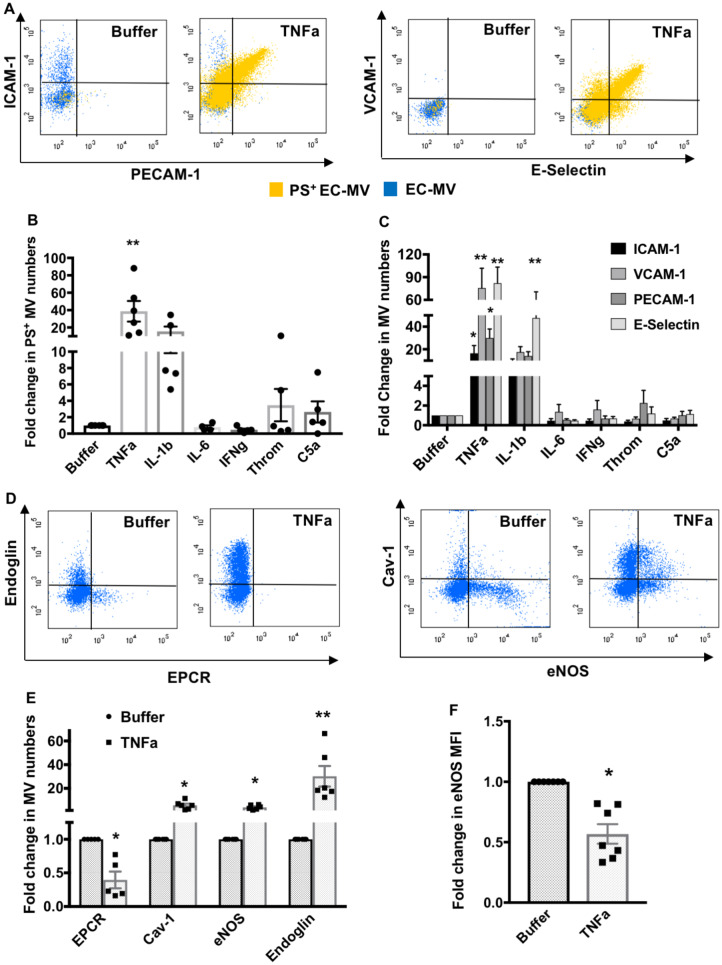
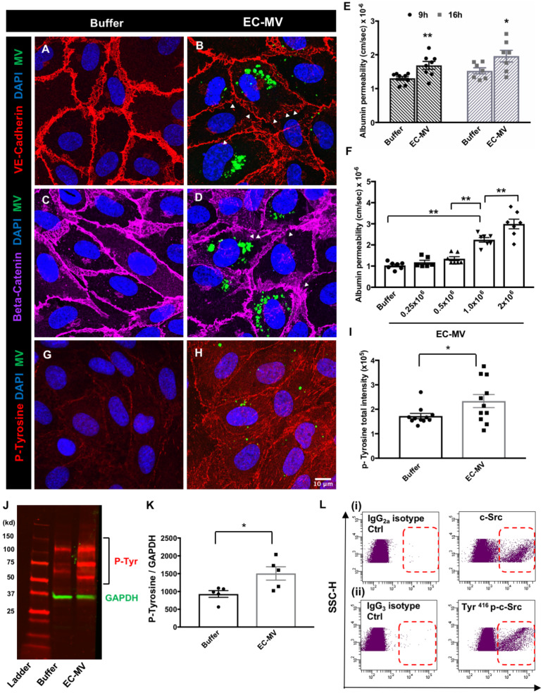
Methods and results: We measured the levels and molecular properties of endothelial-derived MVs (EC-MVs) from mouse plasma following a septic injury elicited by cecal ligation and puncture, as well as those from supernatants of cultured endothelial cells stimulated by inflammatory agents including cytokines, thrombin, and complement 5a. The mouse studies showed that sepsis caused a significant increase in total plasma vesicles and VE-cadherin+ EC-MVs compared to sham control. In cultured ECs, different inflammatory agents caused diverse patterns of EC-MV production and cargo contents. When topically applied to endothelial cells, EC-MVs induced a cytoskeleton-junction response characterized by myosin light chain phosphorylation, contractile fibre reorganization, VE-cadherin phosphorylation, and adherens junction dissociation, functionally measured as increased albumin transendothelial flux and decreased barrier resistance. The endothelial response was coupled with protein tyrosine phosphorylation promoted by MV cargo containing c-Src kinase, whereas MVs produced from c-Src deficient cells did not exert barrier-disrupting effects. Additionally, EC-MVs contribute to endothelial inflammatory injury by promoting neutrophil-endothelium adhesion and release of neutrophil extracellular traps containing citrullinated histones and myeloperoxidase, a response unaltered by c-Src knockdown.

Conclusion: Endothelial-derived microparticles cause endothelial barrier dysfunction by impairing adherens junctions and activating neutrophils. The signalling mechanisms underlying the endothelial cytoskeleton-junction response to EC-MVs involve protein phosphorylation promoted by MV cargo carrying c-Src. However, EC-MV-induced neutrophil activation was not dependent on c-Src.

Keywords: Barrier function; Endothelial cells; Inflammation; Microvesicles; Neutrophils.


Figures

Figure 1
Figure 1
EV levels are increased in mouse plasma during CLP-induced sepsis. (A) Representative NTA tracings show increased concentration of EVs in septic plasma as compared to sham. (i) Merged concentration-size plots of EVs from sham and CLP plasma. The red (CLP) and blue (sham) line graphs show individual concentration values plotted against particle sizes derived from five separate NTA readings. (ii and iii) Intensity-size plots of EVs from mouse plasma that underwent sham surgery or CLP, respectively showing increased particles in CLP plasma. The different shades of green and blue scatter points represent particle tracings from five separate readings. (B) Quantification of NTA analyses shows that the total EV numbers are significantly increased in CLP plasma compared to sham plasma. (C) TEM image of plasma MVs showing particles near the 100 nm size range. Scale bar = 100 nm. Flow cytometry analyses show significantly increased total MV numbers (D) and VE-Cadherin+ MVs in the plasma of CLP mice (E and F). All data are presented as mean ± SEM. *P < 0.05, **P < 0.01 between sham and CLP by Student’s t-test, n = 6–12 animals.
Figure 2
Figure 2
In vitro inflammatory stimulation alters endothelial MV numbers and cargos. (A) Representative flow cytometry dot plots demonstrate increased ICAM-1, PECAM-1, VCAM-1, E-selectin, and PS positive MVs generated from HUVECs treated with TNFα. (B) PS+ EC-MV generation is significantly increased by TNFα treatment (n = 5–6 experiments). (C) TNFα significantly increased ICAM-1+, VCAM-1+, PECAM-1+, and E-selectin+ EC-MV generation, while IL-1β only significantly increased E-selectin+ EC-MV generation from HUVECs (n = 5–10 experiments). **P < 0.01 between buffer and individual inflammatory mediators by one-way ANOVA analysis and Dunnett’s multiple comparison tests. (D and E) Flow cytometry analyses of TNFα-induced EC-MVs containing EPCR+, Cav-1+, eNOS+, and endoglin+ cargos. *P < 0.05, **P < 0.01 between buffer and TNFα by Student’s t-test, n = 5–6 experiments. (F) eNOS expression (median fluorescence intensity) is significantly reduced in EC-MVs induced by TNFα. *P < 0.05 by Student’s t-test, n = 7 experiments. All data are shown as mean ± SEM.
Figure 3
Figure 3
EC-MVs increase endothelial permeability and induce phosphorylation of proteins in recipient cells. (AD) Representative confocal images showing discontinuous adherens junctions in HUVEC monolayers after EC-MV treatment. VE-cadherin (A and B) staining in buffer-treated and MV-treated ECs; (C and D) beta-catenin staining in HUVECs with and without EC-MVs (green dots indicate MVs, arrowheads point to discontinuous junction lining). (E) Albumin permeability through endothelial monolayers is increased after treatment with EC-MVs at 9 and 16 h. *P < 0.05, **P < 0.01 between buffer and EC-MV at 9 h and 16 h, respectively by Student’s t-test, n = 7–8 experiments. (F) Albumin permeability through endothelial monolayers is increased after treatment with increasing concentration of EC-MVs. **P < 0.01 by one-way ANOVA with Tukey’s multiple comparison tests, n = 6–7 experiments. (GI) Representative confocal images showing phospho-tyrosine level is remarkably increased after EC-MV treatment. Scale bar = 10µm and applies to all images. *P < 0.05 by Student’s t-test, n = 11 different confocal z stacks from multiple slides. (J and K) Western blot analysis showing phospho-tyrosine level is significantly increased after EC-MV treatment. *P < 0.05 between control and EC-MV treated cells by Student’s t-test, n = 5 lysates. All data are shown as mean ± SEM. (L) Representative flow cytometry dot plots show the presence of c-Src kinase and Tyr416 phosphorylated c-Src on EC-MVs.
Figure 4
Figure 4
EC-MVs promote stress fibre formation, MLC, and VE-cadherin phosphorylation. (AC) Representative confocal microscopy images and z stack analysis showing increased F-actin stress fibres (arrowheads) in ECs after EC-MV interaction. (DF) The actin-binding protein cortactin is increased and redistributes from the cell periphery to more centralized intracellular locations after EC-MV treatment. Scale bar corresponds to 10µm and applies to all images.**P < 0.01 between buffer and EC-MV treated cells by Student’s t-test, n = 10–12 different confocal z stacks from multiple slides. (G and H) Representative confocal microscopy images showing EC-MV interaction with ECs increase pMLC2 (Thr18/Ser19) which co-stains with F actin (arrowheads). (I) A three-dimensional reconstruction of Panel H reveal MVs fused to the cell membrane as well as inside the cell (arrowheads) at the same focal plane with F-actin stress fibres and pMLC2. (J and K) Representative confocal microscopy images showing EC-MV interaction increases Tyr658 p-VE-Cadherin compared to control cells. (L and M) Representative western blot images and analysis showing increased percentage of phosphorylated MLC2 and VE-cadherin after EC-MV treatment. All data are shown as mean ± SEM. **P < 0.01 by Student’s t-test, n = 8 lysates.
Figure 5
Figure 5
EC-MVs deficient in c-Src have reduced effects on albumin flux, stress fibre formation, MLC, and VE-cadherin phosphorylation. (A) EC-MVs derived from c-Src knockdown HUVECs display negligible c-Src compared to those from scrambled siRNA-treated cells, confirming the efficiency of c-Src knockdown. (B) Western blot image showing knockdown of intracellular c-Src in ECs after transfection with c-Src siRNA. (C) Albumin permeability through endothelial monolayers is increased at 16 h after treatment with scrambled EC-MVs but not by c-Src deficient EC-MVs. *P < 0.05, **P < 0.01 by one-way ANOVA with Tukey’s multiple comparison tests, n = 6 experiments. (DF) Confocal microscopy z stack analysis comparing F-actin levels in endothelial monolayers treated with scrambled vs. Src-deficient EC-MVs. (GI) Comparison of cortactin levels from confocal z stacks in endothelial cells treated with scrambled vs. Src-deficient EC-MVs. *P < 0.05, **P < 0.01 by Student’s t-test, n = 10–18 different confocal z stacks from multiple slides. Representative confocal images showing increased phospho-MLC levels (J and K) and Tyr658 p-VE-Cadherin levels (M and N) in endothelial cells treated with scrambled vs. Src-deficient EC-MVs. Scale bar corresponds to 10µm and applies to all images. (L, O) Western blot images and analysis showing increased percentage of pMLC2 and Tyr658 p-VE-Cadherin in endothelial cells treated with scrambled vs. Src-deficient EC-MVs. Data are shown as mean ± SEM. **P < 0.01 by Student’s t-test, n = 5–8 lysates.
Figure 6
Figure 6
EC-MVs cause activation of neutrophils and endothelial cells. (A and B) Flow cytometry analyses show significantly increased ICAM-1 and VCAM-1 expression on HUVECs after treatment with EC-MVs for 4 h. **P < 0.01 between buffer and EC-MV by Student’s t-test, n = 5 experiments. (C and D) Whole blood flow cytometry analyses show increased levels of CD11b on neutrophil surface after treatment with EC-MVs for 2 h. CD66b was used as a marker of neutrophils. **P < 0.01 by Student’s t-test, n = 7 experiments. (E and F) Representative confocal images and cell adhesion assay showing increased EC-PMN adhesion after EC-MV treatment. **P < 0.01 by Student’s t-test, n = 5 experiments. Arrowheads indicate adherent neutrophils. Scale bar = 20 µm. (G) EC-MV interaction with neutrophils induce NET formation. (Left panel) Unstimulated neutrophil and (right panel) neutrophils stimulated by EC-MVs showing NET formation, arrow points to MVs. Scale bar = 10 µm. (H) Increased percentage of neutrophils undergoing NETosis following treatment with EC-MVs. There was no statistical difference in NET formation between c-Src KD EC-MVs and scrambled EC-MVs. **P < 0.01, ns = not significant by one-way ANOVA analysis, n = 5 experiments. All data are shown as mean ± SEM.
Figure 7
Figure 7
Schematic diagram summarizing the effects of inflammation induced EC-MVs on endothelial and neutrophil activation. Inflammatory injury increases the production of endothelial microvesicles. Src+ EC-MVs disrupt endothelial barrier integrity by increasing acto-myosin contractility through stress fibre formation and increased phosphorylation of MLCK. Src+ EC-MVs increase tyrosine phosphorylation of VE-cadherin and increase albumin permeability through endothelial monolayers. EC-MVs also contribute to vascular injury indirectly by increasing adhesion molecules on ECs and neutrophils to enhance PMN-EC adhesion and by promoting NETosis with release of citrullinated histone H3 and myeloperoxidase. Images of cells were obtained from Smart Servier Medical Art (https://smart.servier.com).

Similar articles

Cited by

References

    1. Yuan SY, Shen Q, Rigor RR, Wu MH.. Neutrophil transmigration, focal adhesion kinase and endothelial barrier function. Microvasc Res 2012;83:82–88. - PMC - PubMed
    1. Yuan SY, Rigor RR.. Regulation of Endothelial Barrier Function. San Rafael, CA: Morgan and Claypool Life Sciences; 2010. - PubMed
    1. Shen Q, Wu MH, Yuan SY.. Endothelial contractile cytoskeleton and microvascular permeability. Cell Health Cytoskelet 2009;2009:43–50. - PMC - PubMed
    1. Raposo G, Stoorvogel W.. Extracellular vesicles: exosomes, microvesicles, and friends. J Cell Biol 2013;200:373–383. - PMC - PubMed
    1. Lo Cicero A, Stahl PD, Raposo G.. Extracellular vesicles shuffling intercellular messages: for good or for bad. Curr Opin Cell Biol 2015;35:69–77. - PubMed

Publication types

MeSH terms