. 2020 May 6;70(10):2045-2053.
doi: 10.1093/cid/ciz600.

A Rare Mutation in SPLUNC1 Affects Bacterial Adherence and Invasion in Meningococcal Disease

Affiliations

A Rare Mutation in SPLUNC1 Affects Bacterial Adherence and Invasion in Meningococcal Disease

Bayarchimeg Mashbat et al. Clin Infect Dis. .

Erratum in

Abstract

Background: Neisseria meningitidis (Nm) is a nasopharyngeal commensal carried by healthy individuals. However, invasive infections occurs in a minority of individuals, with devastating consequences. There is evidence that common polymorphisms are associated with invasive meningococcal disease (IMD), but the contributions of rare variants other than those in the complement system have not been determined.

Methods: We identified familial cases of IMD in the UK meningococcal disease study and the European Union Life-Threatening Infectious Disease Study. Candidate genetic variants were identified by whole-exome sequencing of 2 patients with familial IMD. Candidate variants were further validated by in vitro assays.

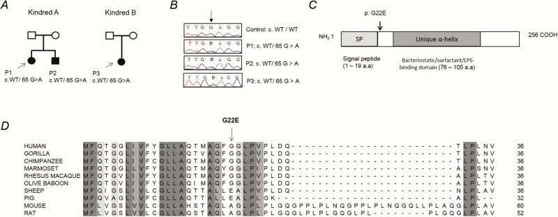
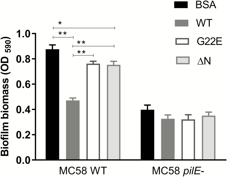
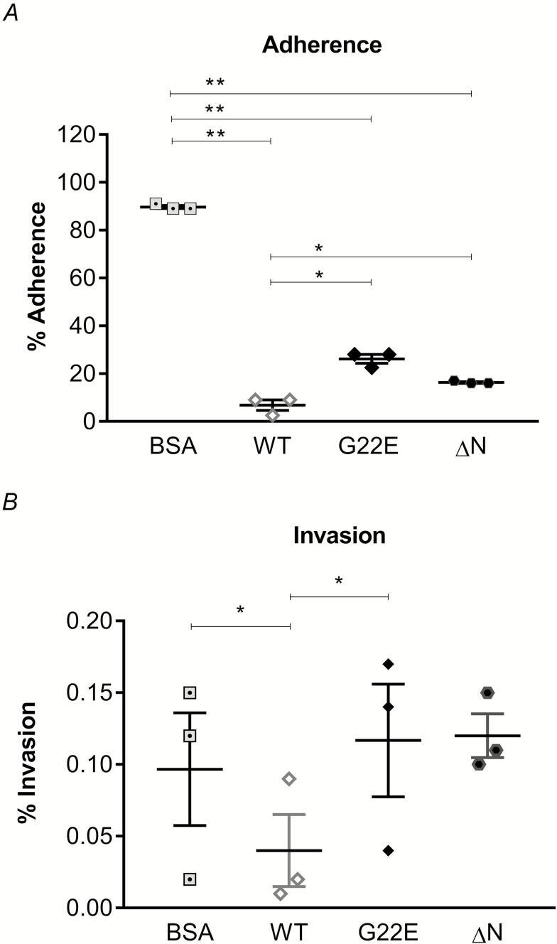
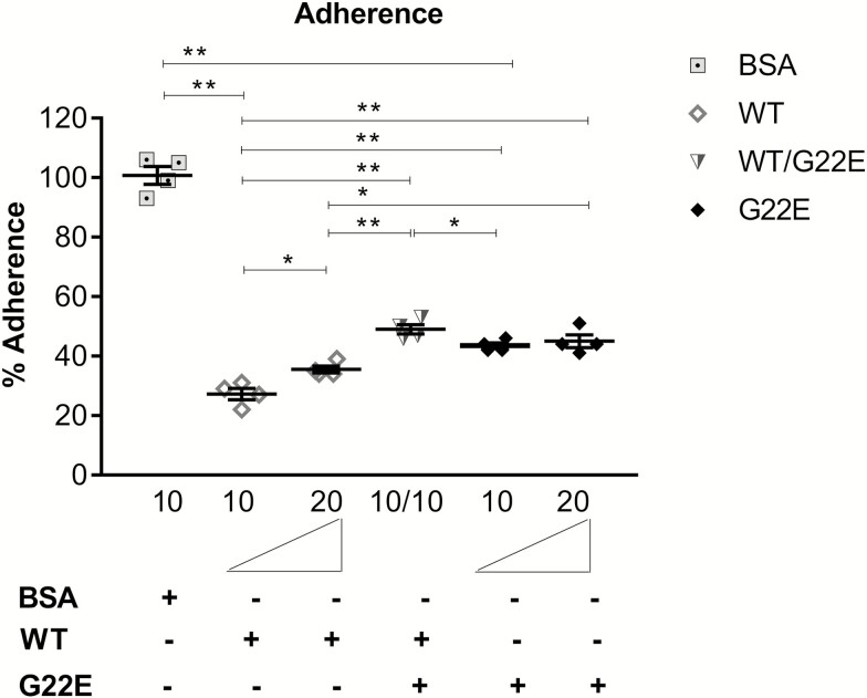
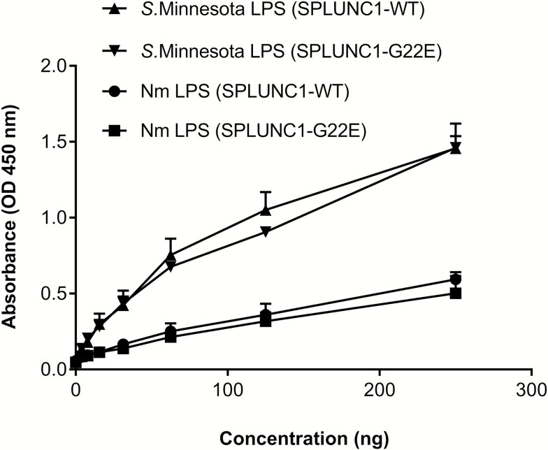
Results: Exomes of 2 siblings with IMD identified a novel heterozygous missense mutation in BPIFA1/SPLUNC1. Sequencing of 186 other nonfamilial cases identified another unrelated IMD patient with the same mutation. SPLUNC1 is an innate immune defense protein expressed in the nasopharyngeal epithelia; however, its role in invasive infections is unknown. In vitro assays demonstrated that recombinant SPLUNC1 protein inhibits biofilm formation by Nm, and impedes Nm adhesion and invasion of human airway cells. The dominant negative mutant recombinant SPLUNC1 (p.G22E) showed reduced antibiofilm activity, increased meningococcal adhesion, and increased invasion of cells, compared with wild-type SPLUNC1.

Conclusions: A mutation in SPLUNC1 affecting mucosal attachment, biofilm formation, and invasion of mucosal epithelial cells is a new genetic cause of meningococcal disease.

Keywords: human genetics; meningococcal disease; mucosal immunity; sepsis; severe infectious disease.


Figures

Figure 1.
Figure 1.
A heterozygous missense BPIFA1/SPLUNC1 mutation identified in invasive meningococcal disease (IMD) patients. A novel missense c.65 G > A, p.G22E SPLUNC1 mutation was found in 3 IMD patients in 2 kindreds. A, Pedigrees of the 2 families’ affected individuals are shaded in black and probands are indicated by arrows. B,SPLUNC1 sequencing from control and patients. Arrow indicates the position of the mutation. C, Schematic representation of the SPLUNC1 protein, with the signal peptide domain (SP) and BPI–alpha/beta homologous domain (BPI-α/β) indicated.) D, Amino acid sequence alignment showing conservation of the N-terminal domain of SPLUNC1 across different species. Abbreviation: COOH, C-terminal domain of proteins; IMD, invasive meningococcal disease LPS, lipopolysaccharide; WT, wild-type.
Figure 2.
Figure 2.
Nm biofilm formation is inhibited by rSPLUNC1. Biofilm biomass formed by WT or pilE-mutant Nm in media supplemented with BSA, WT, mutant G22E, or ΔN mutant rSPLUNC1 proteins are shown on a microtitre plate. Results are expressed as the means ± SEMs from 3 independent experiments. Statistical significance was assessed using a 2-way ANOVA, followed by Tukey’s test. *P < .05; **P < .001. Abbreviations: ΔN, N-terminal deleted; ANOVA, analysis of variance; Nm, Neisseria meningitidis; OD, optical density; pilE, pili deficient; rSPLUNC1, recombinant SPLUNC1; SEM, standard error of the mean; WT, wild-type.
Figure 3.
Figure 3.
Nm growth is not affected by rSPLUNC1. Nm was grown in the presence of WT rSPLUNC1 or BSA for up to (A) 3 hours or (B) 24 hours. The bacterial viability was assessed by counting CFUs at regular intervals during infection. Results are expressed as means ± SDs from at least 3 independent experiments carried out in triplicates. Statistical significance was assessed using the Student’s t-test. Abbreviations: Nm, Neisseria meningitidis; rSPLUNC1, recombinant SPLUNC1; SD, standard deviation; WT, wild-type.
Figure 4.
Figure 4.
Nm adherence and invasion of 16HBE14 is inhibited by rSPLUNC1. The effects of BSA, WT, mutant G22E, or ΔN mutant rSPLUNC1 proteins on (A) Nm adherence or (B) invasion into 16HBE14 cells were assessed. Each condition was carried out in triplicate and the means ± SEMs from 3 independent experiments, following 4 hours incubation, are shown. *P < .05, **P < .001: determined by 1-way ANOVA with Tukey’s test. Abbreviations: ΔN, N-terminal deleted; 16HBE14, human bronchial epithelial cell line; ANOVA, analysis of variance; Nm, Neisseria meningitidis; rSPLUNC1, recombinant SPLUNC1; SEM, standard error of the mean; WT, wild-type.
Figure 5.
Figure 5.
The mutant G22E rSPLUNC1 protein is dominant negative. Bacterial adherence to 16HBE14 was tested in the presence of increasing doses of mutant G22E and/or WT rSPLUNC1. The amounts of recombinant protein used are displayed below the bar. The adherent bacteria were determined by CFU counts. Each condition was carried out in triplicate and the means ± SEMs from 5 independent experiments, following 4 hours adherence, are shown. *P < .05, **P < .001: determined by 1-way ANOVA with Tukey’s test. Abbreviations: 16HBE14, human bronchial epithelial cell line; ANOVA, analysis of variance; rSPLUNC1, recombinant SPLUNC1; SEM, standard error of the mean; WT, wild-type.
Figure 6.
Figure 6.
Both WT and mutant G22E rSPLUNC1 proteins bind LPS. Binding of Nm LPS or Salmonella Minnesota LPS are shown, with increasing doses of either WT or mutant G22E rSPLUNC1 proteins determined by ELISA. All experiments were performed 3 times and results are shown as means ± SDs. Abbreviations: ELISA, enzyme-linked immunosorbent assay; LPS, lipopolysaccharide; Nm, Neisseria meningitidis; OD, optical density; rSPLUNC1, recombinant SPLUNC1; SD, standard deviation; WT, wild-type.

Similar articles

Cited by

References

    1. Yazdankhah SP, Caugant DA. Neisseria meningitidis: an overview of the carriage state. J Med Microbiol 2004; 53:821–32. - PubMed
    1. Virji M. Pathogenic neisseriae: surface modulation, pathogenesis and infection control. Nat Rev Microbiol 2009; 7:274–86. - PubMed
    1. Caugant DA, Maiden MC. Meningococcal carriage and disease–population biology and evolution. Vaccine 2009; 27(Suppl 2):B64–70. - PMC - PubMed
    1. Mace SE. Acute bacterial meningitis. Emerg Med Clin North Am 2008; 26:281–317, viii. - PubMed
    1. Pace D, Pollard AJ. Meningococcal disease: clinical presentation and sequelae. Vaccine 2012; 30(Suppl 2):B3–9. - PubMed

Publication types