

A Subset of Olfactory Sensory Neurons Express Forkhead Box J1-Driven eGFP
- PMID: 31504289
- PMCID: PMC6821233
- DOI: 10.1093/chemse/bjz060
A Subset of Olfactory Sensory Neurons Express Forkhead Box J1-Driven eGFP
Abstract
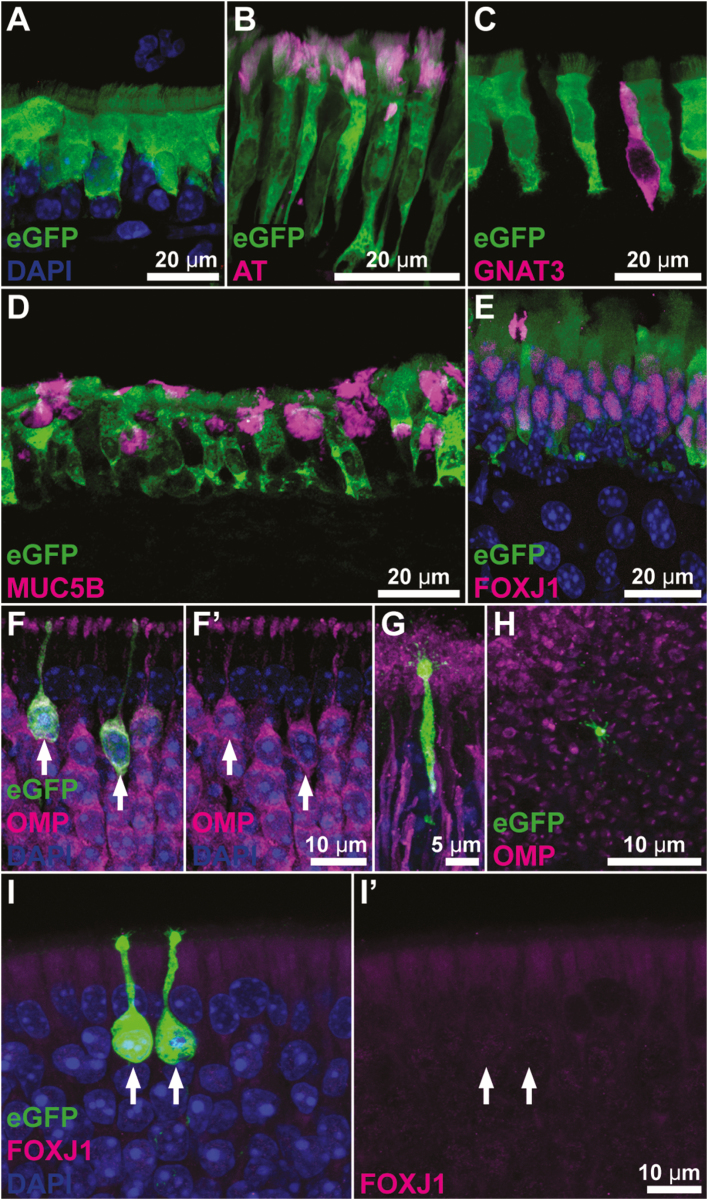
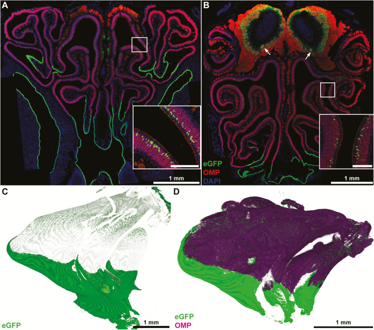
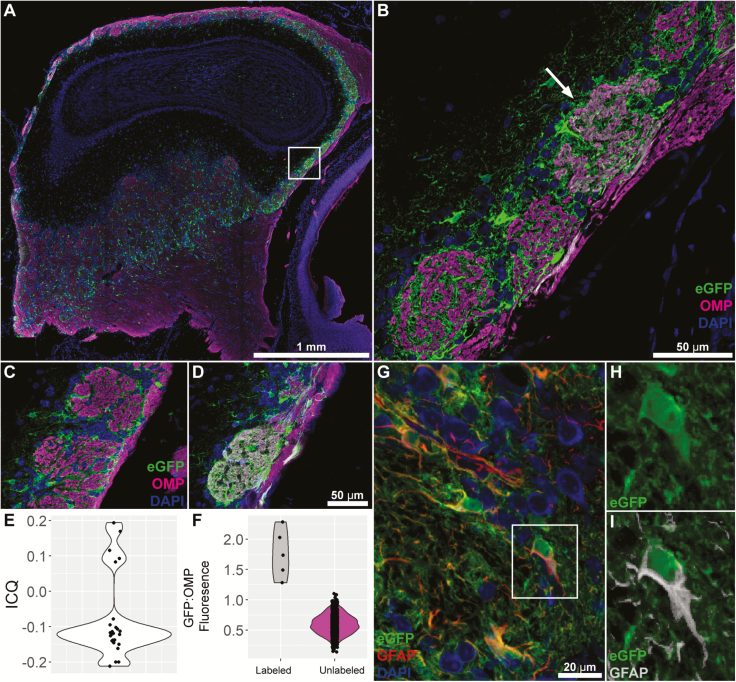
Forkhead box protein J1 (FOXJ1), a member of the forkhead family transcription factors, is a transcriptional regulator of motile ciliogenesis. The nasal respiratory epithelium, but not olfactory epithelium, is lined with FOXJ1-expressing multiciliated epithelial cells with motile cilia. In a transgenic mouse where an enhanced green fluorescent protein (eGFP) transgene is driven by the human FOXJ1 promoter, robust eGFP expression is observed not only in the multiciliated cells of the respiratory epithelium but in a distinctive small subset of olfactory sensory neurons in the olfactory epithelium. These eGFP-positive cells lie at the extreme apical part of the neuronal layer and are most numerous in dorsal-medial regions of olfactory epithelium. Interestingly, we observed a corresponding small number of glomeruli in the olfactory bulb wherein eGFP-labeled axons terminate, suggesting that the population of eGFP+ receptor cells expresses a limited number of olfactory receptors. Similarly, a subset of vomeronasal sensory neurons expresses eGFP and is distributed throughout the full height of the vomeronasal sensory epithelium. In keeping with this broad distribution of labeled vomeronasal receptor cells, eGFP-labeled axons terminate in many glomeruli in both anterior and posterior portions of the accessory olfactory bulb. These findings suggest that Foxj1-driven eGFP marks a specific population of olfactory and vomeronasal sensory neurons, although neither receptor cell population possess motile cilia.
Keywords: FOXJ1; cilia; forkhead transcription factors; olfactory bulb; olfactory receptor neurons.
© The Author(s) 2019. Published by Oxford University Press. All rights reserved. For permissions, please e-mail: journals.permissions@oup.com.
Figures

Similar articles
-
The forkhead transcription factor Foxj1 controls vertebrate olfactory cilia biogenesis and sensory neuron differentiation.PLoS Biol. 2024 Jan 25;22(1):e3002468. doi: 10.1371/journal.pbio.3002468. eCollection 2024 Jan. PLoS Biol. 2024. PMID: 38271330 Free PMC article.
-
Heterogeneous expression of connexin 36 in the olfactory epithelium and glomerular layer of the olfactory bulb.J Comp Neurol. 2003 May 12;459(4):426-39. doi: 10.1002/cne.10617. J Comp Neurol. 2003. PMID: 12687708
-
Olfactory glomeruli are innervated by more than one distinct subset of primary sensory olfactory neurons in mice.J Comp Neurol. 1996 Apr 15;367(4):550-62. doi: 10.1002/(SICI)1096-9861(19960415)367:4<550::AID-CNE6>3.0.CO;2-3. J Comp Neurol. 1996. PMID: 8731225
-
Zonal organization of the mammalian main and accessory olfactory systems.Philos Trans R Soc Lond B Biol Sci. 2000 Dec 29;355(1404):1801-12. doi: 10.1098/rstb.2000.0736. Philos Trans R Soc Lond B Biol Sci. 2000. PMID: 11205342 Free PMC article. Review.
-
Maturation of the Olfactory Sensory Neuron and Its Cilia.Chem Senses. 2020 Dec 5;45(9):805-822. doi: 10.1093/chemse/bjaa070. Chem Senses. 2020. PMID: 33075817 Free PMC article. Review.
Cited by
-
Differential Expression of Mucins in Murine Olfactory Versus Respiratory Epithelium.Chem Senses. 2019 Sep 7;44(7):511-521. doi: 10.1093/chemse/bjz046. Chem Senses. 2019. PMID: 31300812 Free PMC article.
-
Olfactory dysfunction in COVID-19: new insights into the underlying mechanisms.Trends Neurosci. 2023 Jan;46(1):75-90. doi: 10.1016/j.tins.2022.11.003. Epub 2022 Nov 16. Trends Neurosci. 2023. PMID: 36470705 Free PMC article. Review.
-
The role of motile cilia in the development and physiology of the nervous system.Philos Trans R Soc Lond B Biol Sci. 2020 Feb 17;375(1792):20190156. doi: 10.1098/rstb.2019.0156. Epub 2019 Dec 30. Philos Trans R Soc Lond B Biol Sci. 2020. PMID: 31884916 Free PMC article. Review.
References
-
- Blatt EN, Yan XH, Wuerffel MK, Hamilos DL, Brody SL. 1999. Forkhead transcription factor HFH-4 expression is temporally related to ciliogenesis. Am J Respir Cell Mol Biol. 21:168–176. - PubMed
-
- Cahoy JD, Emery B, Kaushal A, Foo LC, Zamanian JL, Christopherson KS, Xing Y, Lubischer JL, Krieg PA, Krupenko SA, et al. . 2008. A transcriptome database for astrocytes, neurons, and oligodendrocytes: a new resource for understanding brain development and function. J Neurosci. 28:264–278. - PMC - PubMed
Publication types
MeSH terms
Substances
Grants and funding
LinkOut - more resources
Full Text Sources
Molecular Biology Databases
