. 2019 Oct 15;70(19):5271-5285.
doi: 10.1093/jxb/erz275.

The pyrenoidal linker protein EPYC1 phase separates with hybrid Arabidopsis-Chlamydomonas Rubisco through interactions with the algal Rubisco small subunit

Affiliations

The pyrenoidal linker protein EPYC1 phase separates with hybrid Arabidopsis-Chlamydomonas Rubisco through interactions with the algal Rubisco small subunit

Nicky Atkinson et al. J Exp Bot. .

Abstract

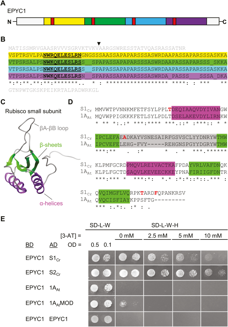
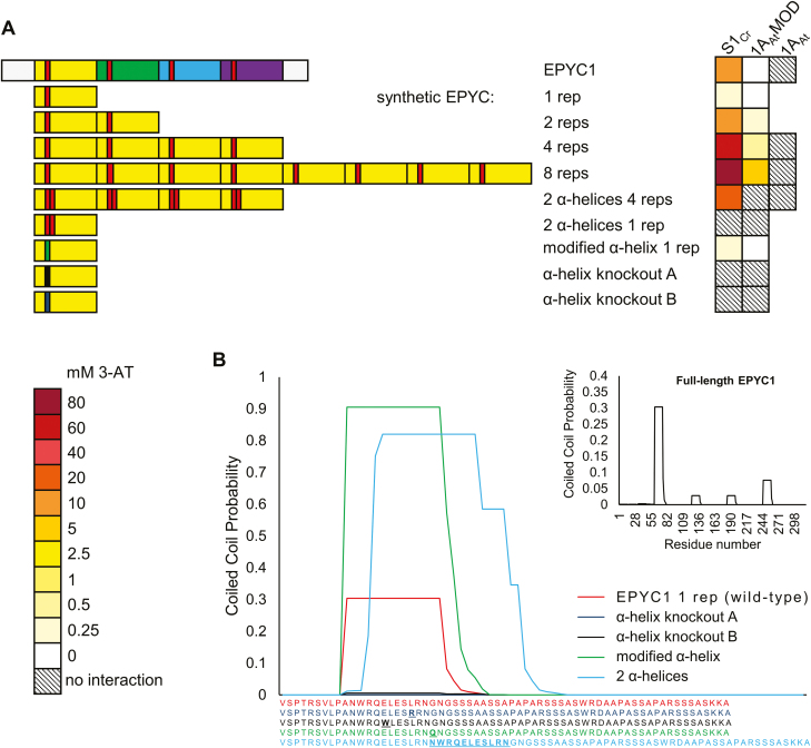
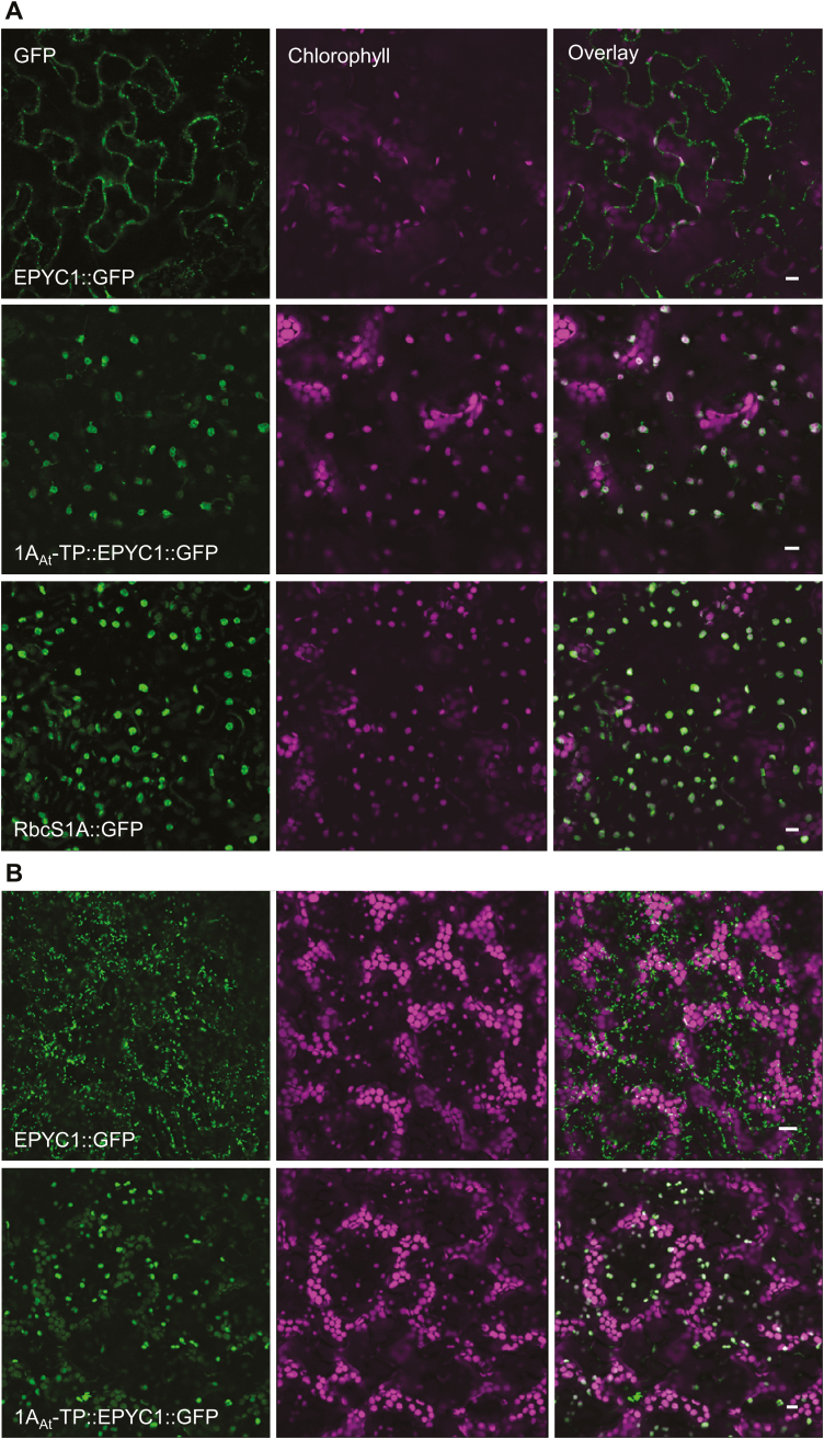
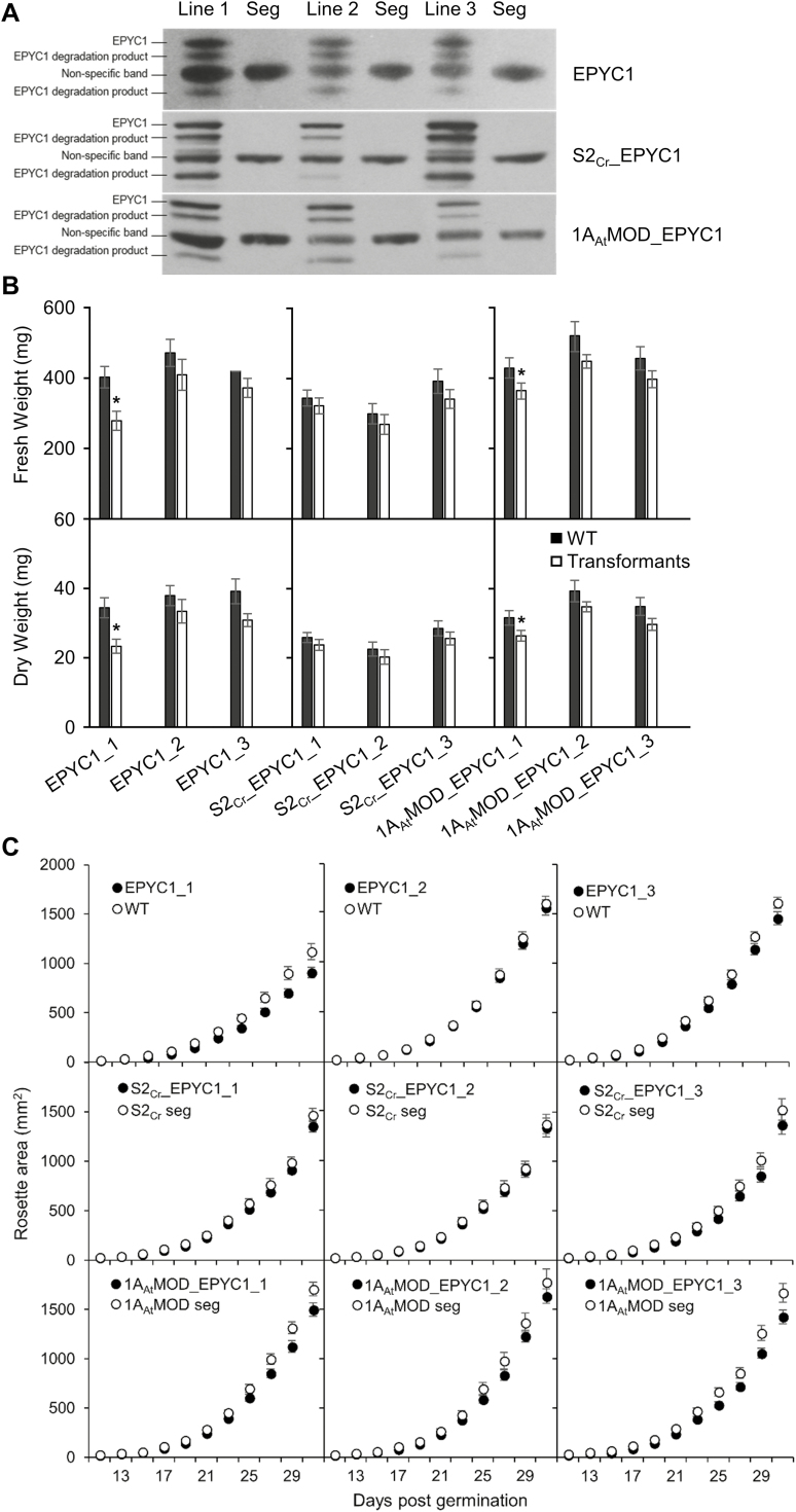
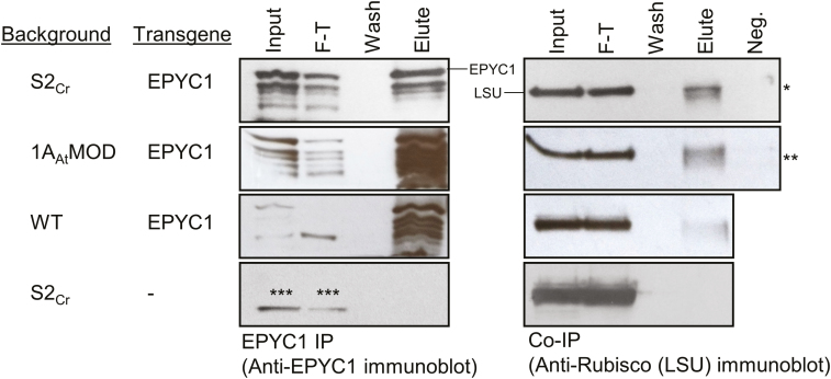
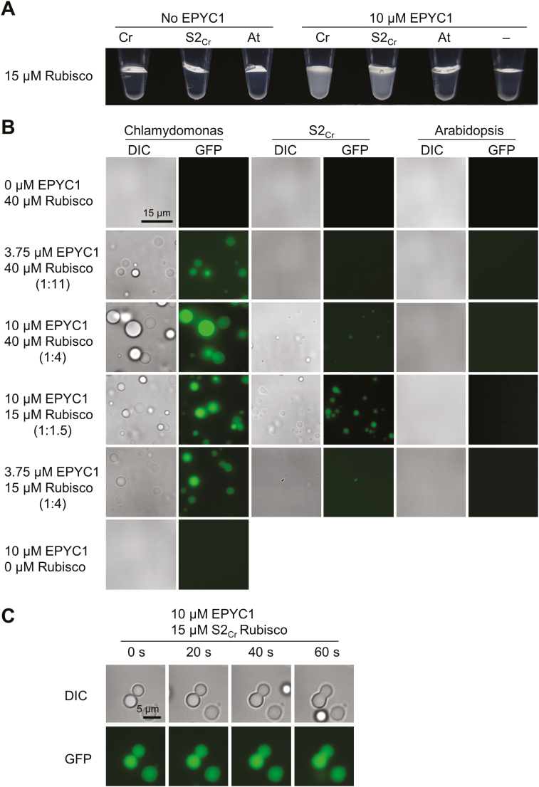
Photosynthetic efficiencies in plants are restricted by the CO2-fixing enzyme Rubisco but could be enhanced by introducing a CO2-concentrating mechanism (CCM) from green algae, such as Chlamydomonas reinhardtii (hereafter Chlamydomonas). A key feature of the algal CCM is aggregation of Rubisco in the pyrenoid, a liquid-like organelle in the chloroplast. Here we have used a yeast two-hybrid system and higher plants to investigate the protein-protein interaction between Rubisco and essential pyrenoid component 1 (EPYC1), a linker protein required for Rubisco aggregation. We showed that EPYC1 interacts with the small subunit of Rubisco (SSU) from Chlamydomonas and that EPYC1 has at least five SSU interaction sites. Interaction is crucially dependent on the two surface-exposed α-helices of the Chlamydomonas SSU. EPYC1 could be localized to the chloroplast in higher plants and was not detrimental to growth when expressed stably in Arabidopsis with or without a Chlamydomonas SSU. Although EPYC1 interacted with Rubisco in planta, EPYC1 was a target for proteolytic degradation. Plants expressing EPYC1 did not show obvious evidence of Rubisco aggregation. Nevertheless, hybrid Arabidopsis Rubisco containing the Chlamydomonas SSU could phase separate into liquid droplets with purified EPYC1 in vitro, providing the first evidence of pyrenoid-like aggregation for Rubisco derived from a higher plant.

Keywords: Arabidopsis thaliana; Chlamydomonas reinhardtii; Nicotiana benthamiana; CO2-concentrating mechanism; chloroplast; photosynthesis; pyrenoid.


Figures

Fig. 1.
Fig. 1.
EPYC1 interacts with the Rubisco small subunit. (A and B) EPYC1 consists of four near identical repeat regions (highlighted in yellow, green, blue, and purple), each containing a predicted α-helix (red, underlined). The putative cleavage site of the chloroplastic transit peptide between 26 (V) and 27 (A) is indicated. The N- and C- termini are shown in grey. (C) The predicted model of the small subunit of Rubisco (SSU; 1AAt shown) has similar features in algae and higher plants, including four β-sheet (green) and two α-helical (purple) regions. (D) Amino acid alignments of mature Arabidopsis SSU 1A (1AAt) and Chlamydomonas SSU 1 (S1Cr). Residues in red indicate the four amino acids that differ between the two Chlamydomonas SSUs, S1Cr and S2Cr (T/S, A/S, T/S, F/W). (E) Yeast two-hybrid interactions with EPYC1. Interaction strength is demonstrated by growth on increasing concentrations of the inhibitor 3-AT. Abbreviations: 3-AT, 3-amino-1,2,4-triazole; BD, binding domain; AD, activation domain; 1AAtMOD, modified 1AAt carrying the two α-helical regions from Chlamydomonas; SD-L-W, yeast synthetic minimal medium (SD medium) lacking leucine (L) and tryptophan (W); SD-L-W-H, SD medium lacking L, W, and histidine (H). See Supplementary Fig. S1 for Y2H additional controls.
Fig. 2.
Fig. 2.
EPYC1 requires the α-helices of the Chlamydomonas SSU for interaction. Interaction is shown between EPYC1 and modified versions of 1AAt (top), in which different components (see Fig. 1C, D) have been switched for those from Chlamydomonas S1Cr (bottom). The heat map indicates interaction strength measured with yeast two-hybrid assays by the capacity for growth on increasing concentrations of 3-AT (mM). See Supplementary Fig. S2 for SSU sequences and Supplementary Fig. S3A for raw Y2H image data.
Fig. 3.
Fig. 3.
EPYC1 repeat regions contribute to interaction with the Rubisco small subunit. (A) Decreasing the number of EPYC1 repeat regions reduced the strength of interaction with S1Cr. (B) The predicted α-helical region in each repeat (red) is important for interaction with S1Cr. These were eliminated by matutation to seven alanines in each of the different EPYC1 variants. The heat map indicates interaction strength measured with yeast two-hybrid assays by the capacity for growth on increasing concentrations of 3-AT (mM). See Supplementary Fig. S3B and C for raw Y2H image data.
Fig. 4.
Fig. 4.
EPYC1 can be modified to increase the interaction strength with SSUs. (A) Synthetic variants of EPYC1 are based on the first repeat regions (yellow) and the predicted α-helix (red). The heat map indicates interaction strength measured with yeast two-hybrid assays by the capacity for growth on increasing concentrations of 3-AT (mM). See Supplementary Fig. S3D for raw Y2H image data. (B) Predicted coiled-coil domain strengths for synthetic variants of the first repeat region of EPYC1 using the PCOILS bioinformatic tool. Matching colour-coded amino acid sequences are shown below, with residues that differ from the wild-type sequence shown in bold. The inlay shows the prediction for full-length EPYC1.
Fig. 5.
Fig. 5.
Expression of GFP-fused EPYC1 with and without the 1AAt chloroplastic transit peptide (1AAt-TP). (A) The constructs were expressed transiently in Nicotiana benthamiana, alongside a GFP-fused Arabidopsis 1A small subunit of Rubisco (RbcS1A::GFP). (B) Stable expression in Arabidopsis. Green and purple signals are GFP and chlorophyll autofluoresence, respectively. Overlapping signals are white. Scale bar=10 µm.
Fig. 6.
Fig. 6.
Growth of Arabidopsis plant lines expressing EPYC1 fused with the 1AAt-TP in either the wild-type (WT), S2Cr, or the 1AAtMOD background. (A) Immunoblots show the relative EPYC1 expression levels in three independently transformed lines (T3) per background, compared with their corresponding segregants (seg) lacking EPYC1. (B) Plants were harvested at 31 d, and the fresh (FW) and dry (DW) weights were measured. (C) Rosette growth of the nine transformed lines. Values are the means ±SE of measurements made on 12 (FW, DW) or 16 (growth assays) rosettes. Asterisks indicate significant difference in FW or DW between transformed lines and segregants (P<0.05) as determined by Student’s paired sample t-tests.
Fig. 7.
Fig. 7.
Co-immunoprecipitation of Rubisco with EPYC1. EPYC1 interacts with Rubisco in transgenic S2Cr, 1AAtMOD, and wild-type (WT) lines expressing EPYC1 fused with the 1AAt-TP. Co-immunoprecipitation was performed using Protein-A coated beads that had been cross-linked to anti-EPYC1 antibody. The input, flow-through (F-T), fourth wash, and boiling elute were run on an SDS–PAGE gel, transferred to a nitrocellulose membrane, and probed with either anti-Rubisco or anti-EPYC1 antibody. Negative controls (Neg.) were carried out by replacing the anti-EPYC1 antibody on the Protein A beads with either anti-HA antibody (*) or no antibody (**) and proceeding with IP as before (only the eluted sample is shown). Asterisks (***) indicate a non-specific band observed with the anti-EPYC1 antibody in all samples including the control line not expressing EPYC1 (S2Cr).
Fig. 8.
Fig. 8.
Hybrid Arabidopsis Rubisco carrying a Chlamydomonas small subunit is able to phase separate and form liquid droplets with EPYC1. (A) Addition of EPYC1 to Rubisco results in turbidity in Chlamydomonas (Cr) and hybrid (S2Cr), but not Arabidopsis (At) Rubisco (shown at ~3 min after mixing at room temperature). (B) The turbidity is caused by the formation of spherical droplets. Fluoresence in samples containing EPYC1 is due to the inclusion of EPYC1::GFP (0.25 µM). (C) Droplets from S2Cr Rubisco and EPYC1 fuse by coalescence. See Supplementary Fig. S11 for droplet sedimentation analysis.

Comment in

Similar articles

Cited by

References

    1. Aigner H, Wilson RH, Bracher A, Calisse L, Bhat JY, Hartl FU, Hayer-Hartl M. 2017. Plant RuBisCo assembly in E. coli with five chloroplast chaperones including BSD2. Science 358, 1272–1278. - PubMed
    1. Atkinson N, Feike D, Mackinder LC, Meyer MT, Griffiths H, Jonikas MC, Smith AM, McCormick AJ. 2016. Introducing an algal carbon-concentrating mechanism into higher plants: location and incorporation of key components. Plant Biotechnology Journal 14, 1302–1315. - PMC - PubMed
    1. Atkinson N, Leitão N, Orr DJ, Meyer MT, Carmo-Silva E, Griffiths H, Smith AM, McCormick AJ. 2017. Rubisco small subunits from the unicellular green alga Chlamydomonas complement Rubisco-deficient mutants of Arabidopsis. New Phytologist 214, 655–667. - PMC - PubMed
    1. Badger MR, Andrews TJ, Whitney SM, Ludwig M, Yellowlees DC, Leggat W, Price GD. 1998. The diversity and coevolution of Rubisco, plastids, pyrenoids, and chloroplast-based CO2-concentrating mechanisms in algae. Canadian Journal of Botany 76, 1052–1071.
    1. Bathellier C, Tcherkez G, Lorimer GH, Farquhar GD. 2018. Rubisco is not really so bad. Plant, Cell & Environment 41, 705–716. - PubMed

Publication types

MeSH terms

Substances