. 2019 Oct 10;47(18):9871-9887.
doi: 10.1093/nar/gkz728.

RsaC sRNA modulates the oxidative stress response of Staphylococcus aureus during manganese starvation

Affiliations

RsaC sRNA modulates the oxidative stress response of Staphylococcus aureus during manganese starvation

David Lalaouna et al. Nucleic Acids Res. .

Abstract

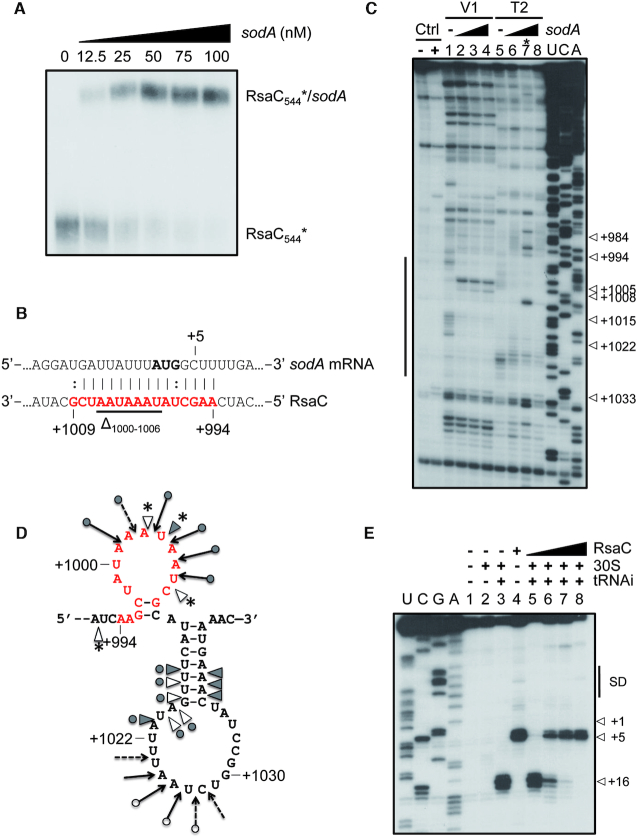
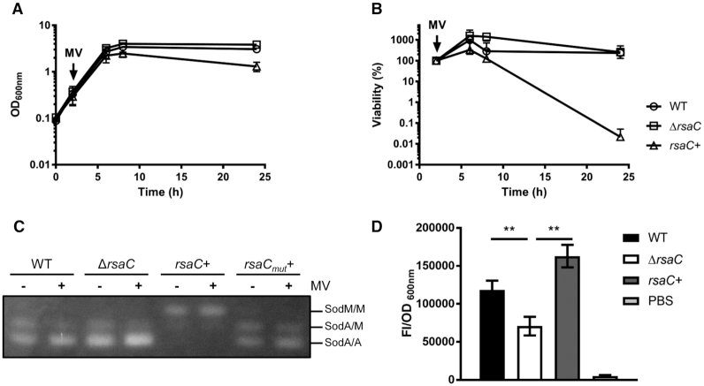
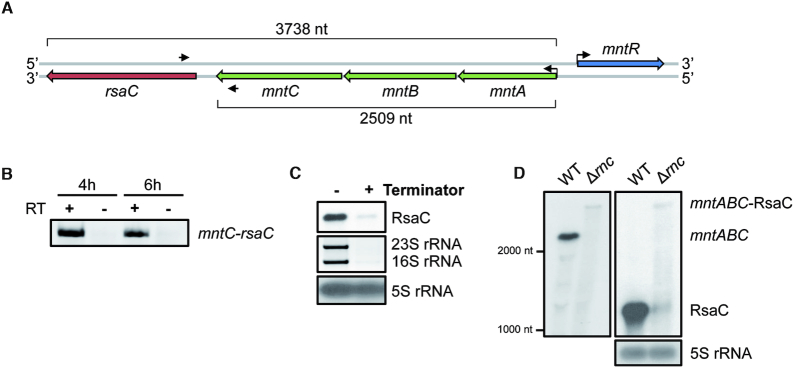
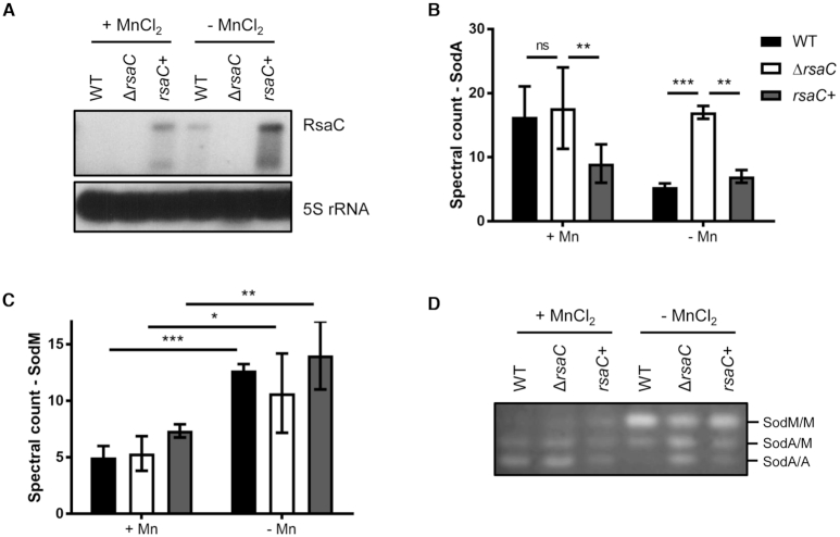
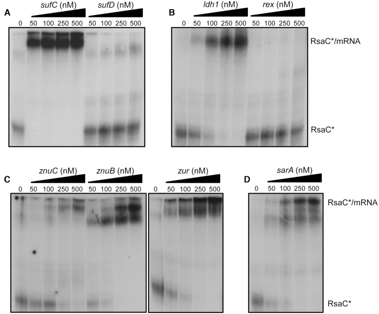
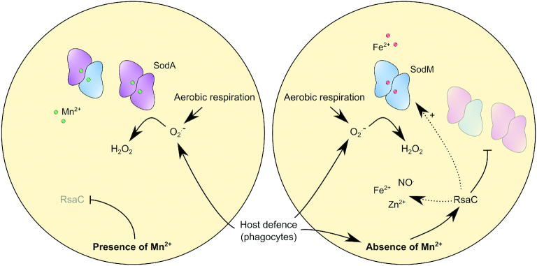
The human opportunistic pathogen Staphylococcus aureus produces numerous small regulatory RNAs (sRNAs) for which functions are still poorly understood. Here, we focused on an atypical and large sRNA called RsaC. Its length varies between different isolates due to the presence of repeated sequences at the 5' end while its 3' part is structurally independent and highly conserved. Using MS2-affinity purification coupled with RNA sequencing (MAPS) and quantitative differential proteomics, sodA mRNA was identified as a primary target of RsaC sRNA. SodA is a Mn-dependent superoxide dismutase involved in oxidative stress response. Remarkably, rsaC gene is co-transcribed with the major manganese ABC transporter MntABC and, consequently, RsaC is mainly produced in response to Mn starvation. This 3'UTR-derived sRNA is released from mntABC-RsaC precursor after cleavage by RNase III. The mature and stable form of RsaC inhibits the synthesis of the Mn-containing enzyme SodA synthesis and favors the oxidative stress response mediated by SodM, an alternative SOD enzyme using either Mn or Fe as co-factor. In addition, other putative targets of RsaC are involved in oxidative stress (ROS and NOS) and metal homeostasis (Fe and Zn). Consequently, RsaC may balance two interconnected defensive responses, i.e. oxidative stress and metal-dependent nutritional immunity.


Figures

Figure 1.
Figure 1.
RsaC is a large and highly structured sRNA. (A) Northern blot analysis of RsaC sRNA in three different S. aureus strains (HG001, MW2 and Newman). Total RNA was extracted after 2, 4 and 6 h of growth in BHI at 37°C. 5S rRNA was used as loading control. Data are representative of three independent experiments. (B) Secondary structure model of RsaC. The structure of RsaC (from HG001 strain) was mapped using selective 2′-hydroxyl acylation by benzol cyanide and the reactivity was analyzed by primer extension (SHAPE). Data were analyzed, and each modification was quantified using QuSHAPE software. The secondary structure was drawn using RNA structure and VARNA considering the reactivity of each ribose. The regions, which were not mapped, are shown in blue. The color code for each reactivity is given on the right side of the secondary structure model, red is for the highest reactivity while green is for the lowest reactivity. A black arrow indicates the +1 of RsaC544.
Figure 2.
Figure 2.
RsaC binds to sodA mRNA and blocks its translation. (A) Gel retardation assays using RsaC544 sRNA and full-length sodA mRNA. 5′ end-radiolabeled RsaC544 (*) was incubated with increasing concentrations of sodA mRNA (from 0 to 100 nM). (B) The RsaC:sodA pairing site predicted using IntaRNA software is indicated in red. The AUG start codon is in bold. Deleted nucleotides (Δ1000–1006) in RsaCmut sequence are underlined. (C) Footprint of sodA mRNA on RsaC. Incubation controls (Ctrl) were performed in absence of RNases on RsaC either free (–) or bound to sodA (50 nM, +). Reactions with either RNase V1 (V1) or RNase T2 (T2) were performed with RsaC (50 nM) in absence (lanes 1 and 5) or in presence of increasing concentrations of sodA mRNA (lanes 2–4 and 6–8; 75, 150 or 300 nM, respectively). U, C, A: sequencing ladders. *Lane 7 should not be considered due to a technical glitch. On the left side, a bar denotes changes in cleavages as the result of sodA binding. (D) Schematic representation of sodA pairing site on RsaC sRNA revealed by footprinting assays. RNase V1 cleavages are denoted by grey triangles for strong cut and empty triangles for moderate cut. RNase T2 cleavages are shown by filled arrows for strong cut and dashed arrows for weak cut. New RNase V1 cleavages are labelled by stars. The predicted pairing site by IntaRNA software is indicated in red. (E) Toe-printing assay monitoring the effect of RsaC on the formation of the ternary ribosomal initiation complex comprising S. aureus 30S subunit, initiator tRNAfMet and sodA mRNA (50 nM). Lanes 1–4: incubation controls (lane 1: sodA mRNA alone; lane 2: sodA mRNA with 30S subunit; lane 3: sodA mRNA with 30S subunit and initiator tRNAfMet (tRNAi); lane 4: sodA mRNA with RsaC); lanes 5–8: formation of the initiation complex in presence of increasing concentrations of RsaC (25, 50, 100, 400 nM). U, C, G, A: sequencing ladders; SD: Shine-Dalgarno sequence; +1: start codon; +5: RT stop induced by RsaC binding; +16: toe-printing signal. Data are representative of two independent experiments.
Figure 3.
Figure 3.
RsaC is involved in oxidative stress response via the repression of SOD activity. (A) Growth monitoring of WT/pCN51-P3 (○), ΔrsaC/pCN51-P3 (□) and ΔrsaC/pCN51-P3-RsaC1116 (rsaC+) (Δ) upon internal superoxide stress (10 mM methyl viologen (MV)). MV was added after 2h of growth. (B) Viability assays of WT/pCN51-P3 (○), ΔrsaC/pCN51-P3 (□) and ΔrsaC/pCN51-P3-RsaC1116 (rsaC+) (Δ) grown in BHI and challenged with 10 mM MV at early-exponential phase (2 h). Samples were taken thereafter, and viability was determined by performing viable counts on GP plates containing 5 μg/ml erythromycin. All data shown represent mean ± SD of three independent experiments. (C) Monitoring of SOD activity in WT/pCN51-P3 (WT), ΔrsaC/pCN51-P3 (ΔrsaC), ΔrsaC/pCN51-P3-RsaC1116 (rsaC+) and ΔrsaC/pCN51-P3-RsaC1116mut (rsaCmut+) after 8h of growth (in presence or absence of MV). SOD activity was determined by negative staining on native polyacrylamide gel (NBT/riboflavin method). Results are representative of two independent experiments. (D) Measurement of reactive oxygen species (ROS) accumulation using fluorescence assays (after 6 h of growth in TSB) in WT/pCN51-P3 (black), ΔrsaC/pCN51-P3 (white) and ΔrsaC/pCN51-P3-RsaC1116 (rsaC+) (gray). PBS buffer (light grey) was used as a control. Data represent mean ± SD of three independent experiments. Statistical analysis with ANOVA, ** P<0.005. Strains and plasmid constructs are given in Supplementary Table S1.
Figure 4.
Figure 4.
rsaC is part of the polycistronic operon mntABC and is released after RNase III cleavage. (A) Genomic context of mntABC operon. Oligonucleotides used for RT-PCR are indicated with black arrows. (B) RT-PCR using oligonucleotides in both mntC and rsaC genes and total RNA extracted after 4 or 6 h of growth in BHI medium. Same experiment was performed without RT enzyme (–) as a control. (C) Determination of the 5′ end status of RsaC sRNA. 10 μg of total RNA extracted after 6 h of growth in BHI medium were treated with the Terminator™ 5′-Phosphate-Dependent Exonuclease. 16S and 23 rRNAs were used as a positive control and 5S rRNA as a negative control. (D) Northern blot analysis of mntABC-RsaC transcript in wild type strain (WT, HG001) and the isogenic RNase III mutant (Δrnc) strain. Total RNA was extracted after 6 h of growth in BHI at 37°C. We used a probe targeting mntA (left panel) or RsaC (right panel). 5S rRNA was used as loading control. Data are representative of three independent experiments.
Figure 5.
Figure 5.
RsaC is involved in Mn homeostasis. (A) Northern blot analysis of RsaC level in WT/pCN51-P3 (WT), ΔrsaC/pCN51-P3 (ΔrsaC) and ΔrsaC/pCN51-P3-RsaC1116 (rsaC+) strains. Total RNA was extracted after 6 h of growth in BHI-chelex supplemented or not with 25 μM MnCl2 at 37°C. 5S rRNA was used as loading control. When RsaC is highly produced, a shorter form is observed. Data are representative of three independent experiments. Differential proteomic analysis of SodA (B) and SodM (C) in presence or absence of 25 μM MnCl2 (BHI-chelex). Data represent spectral count mean ± SD of three independent experiments. Statistical analysis is based on a negative-binomial test using an edgeR GLM regression through R, *P< 0.05; **P< 0.005; ***P< 0.0005; ns: not significant. (D) Monitoring of SOD activity in WT/pCN51-P3 (WT), ΔrsaC/pCN51-P3 (ΔrsaC) and ΔrsaC/pCN51-P3-RsaC1116 (rsaC+) after 6h of growth (in BHI-chelex ± 25 μM MnCl2). SOD activity was determined by negative staining on native polyacrylamide gel (NBT/riboflavin method). Results are representative of three independent experiments. Strains and plasmid constructs are given in Supplementary Table S1.
Figure 6.
Figure 6.
RsaC potentially regulates additional mRNA targets involved in oxidative stress response and metal homeostasis. Gel retardation assays using RsaC544 sRNA and several mRNA targets identified by MAPS and/or proteomic analysis. 5′ end-radiolabeled RsaC544 (*) was incubated with increasing concentrations of (A) sufC and sufD, (B) ldh1 and rex, (C) znuC, znuB and zur, and (D) sarA mRNA (from 0 to 500 nM). Data are representative of two independent experiments. All transcripts have been described in Material and Methods.
Figure 7.
Figure 7.
RsaC sRNA modulates oxidative stress response during Mn starvation. SodA is a Mn-dependent superoxide dismutase which is crucial to response to oxidative stress generated by aerobic respiration or by host defence cells (e.g. macrophages, neutrophils). In absence of Mn, RsaC is highly produced and negatively regulates sodA mRNA translation. It enables to spare Mn for essential Mn-containing proteins and avoids the synthesis of a non-functional enzyme. SodM, an alternative SOD enzyme using Fe as co-factor, replaces SodA to re-establish the ROS detoxification pathway. RsaC may indirectly activate SodM synthesis via the repression of the transcriptional regulator SarA. In addition, RsaC could have a broader role in oxidative stress response, notably through the regulation of Fe and Zn homeostasis (transport and storage) and NO. resistance.

Similar articles

Cited by

References

    1. Flannagan R.S., Heit B., Heinrichs D.E.. Antimicrobial mechanisms of macrophages and the immune evasion strategies of Staphylococcus aureus. Pathogens. 2015; 4:826–868. - PMC - PubMed
    1. Boe D.M., Curtis B.J., Chen M.M., Ippolito J.A., Kovacs E.J.. Extracellular traps and macrophages: new roles for the versatile phagocyte. J. Leukocyte Biol. 2015; 97:1023–1035. - PMC - PubMed
    1. Brinkmann V., Zychlinsky A.. Neutrophil extracellular traps: is immunity the second function of chromatin. J. Cell biol. 2012; 198:773–783. - PMC - PubMed
    1. Imlay J.A. Pathways of oxidative damage. Annu. Rev. microbiol. 2003; 57:395–418. - PubMed
    1. Kehl-Fie T.E., Chitayat S., Hood M.I., Damo S., Restrepo N., Garcia C., Munro K.A., Chazin W.J., Skaar E.P.. Nutrient metal sequestration by calprotectin inhibits bacterial superoxide defense, enhancing neutrophil killing of Staphylococcus aureus. Cell Host Microbe. 2011; 10:158–164. - PMC - PubMed

Publication types

MeSH terms