. 2019 Oct 10;47(18):9888-9901.
doi: 10.1093/nar/gkz732.

Regulation of La/SSB-dependent viral gene expression by pre-tRNA 3' trailer-derived tRNA fragments

Affiliations

Regulation of La/SSB-dependent viral gene expression by pre-tRNA 3' trailer-derived tRNA fragments

Hee Cho et al. Nucleic Acids Res. .

Abstract

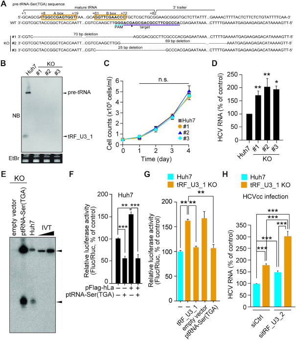
tRNA-derived RNA fragments (tRFs) have emerged as a new class of functional RNAs implicated in cancer, metabolic and neurological disorders, and viral infection. Yet our understanding of their biogenesis and functions remains limited. In the present study, through analysis of small RNA profile we have identified a distinct set of tRFs derived from pre-tRNA 3' trailers in the hepatocellular carcinoma cell line Huh7. Among those tRFs, tRF_U3_1, which is a 19-nucleotide-long chr10.tRNA2-Ser(TGA)-derived trailer, was expressed most abundantly in both Huh7 and cancerous liver tissues, being present primarily in the cytoplasm. We show that genetic loss of tRF_U3_1 does not affect cell growth and it is not involved in Ago2-mediated gene silencing. Using La/SSB knockout Huh7 cell lines, we demonstrate that this nuclear-cytoplasmic shuttling protein directly binds to the 3' U-tail of tRF_U3_1 and other abundantly expressed trailers and plays a critical role in their stable cytoplasmic accumulation. The pre-tRNA trailer-derived tRFs capable of sequestering the limiting amounts of La/SSB in the cytoplasm rendered cells resistant to various RNA viruses, which usurp La/SSB with RNA chaperone activity for their gene expression. Collectively, our results establish the trailer-derived tRF-La/SSB interface, regulating viral gene expression.


Figures

Figure 1.
Figure 1.
Profiles of pre-tRNA 3′ trailer-derived tRFs in the HCC cell line Huh7. (A) Pie chart representing the proportion of small RNAs in Huh7 cells (based on average RPM values from the two small RNA datasets GSM3666020 and GSM3666021). mt, mitochondria. (B) Proportion of tRFs (detected in the above-described datasets) derived from the selected isodecoder tRNA groups (top) and number of tRNA-coding genes for the indicated tRFs (bottom). tRFs in Huh7 cells were derived from a total of 54 isodecoder tRNA-coding genes (out of >270 isodecoder genes in human genome). After analysis of the proportion of tRFs for each isodecoder tRNA group, the ones showing > 0.1 in at least one proportion of each tRF family are presented. (C) The trailer-derived tRF_U3 family tRFs (> 100 average RPM values) sorted in descending order according to their read numbers. The enzymes involved in pre-tRNA processing are indicated. (D) Northern blot analysis for tRF_U3_1. Probes specific to the indicated trailers were used for analysis of total RNA (20 μg) from the indicated cell lines and non-cancerous human liver tissues along with synthetic tRF_U3_1 (0.1 ng). (E) Box-and-whisker plots showing tRF_U3_1 levels (read counts normalized using DESeq2) in human liver tissues [NC, normal (n = 22); C, cancer (n = 14)]. Middle bar, median; box, inter-quartile range (25–75%); bars extend to 1.5× the interquartile range. (F) Northern blot and immunoblot analyses of total RNA and proteins from the indicated subcellular fractions derived from an equal number (4.5 × 10⁶) of Huh7 cells. Cellular fractionation was verified by immunoblotting (IB) for the indicated subcellular organelle markers. Nucl, nuclear fraction; Cyto, cytoplasmic fraction. (G) Major tRF_U3_1 isomers. (H) U-tail length variance in the trailer-derived tRFs in Huh7 cells.
Figure 2.
Figure 2.
Identification of La/SSB as a tRF_U3_1-interacting protein. (A) Cellular proteins interacting with tRF_U3_1 were pulled down from Huh7 cell lysates using 5′-biotin-labeled tRF_U3_1 immobilized onto streptavidin beads, resolved by SDS-PAGE, and visualized by Coomassie blue staining. (B) A PAR-CLIP experiment was carried out to verify the specific interaction of tRF_U3_1 with La/SSB in the Huh7-derived cell line R-1, in which an HCV subgenomic replicon stably replicates. From the La/SSB crosslinked to RNA (marked with an arrowhead), RNA was recovered for TaqMan RT-qPCR quantification of the indicated small RNAs. ND, not detected. (C) Interaction between tRF_U3_1 and La/SSB assessed by a pull-down assay followed by immunoblotting analysis. Pull-down experiments were performed in the absence (−) or presence (+) of the indicated competitors (10-fold molar excess of 5′-biotin-labeled tRF_U3_1). (D) EMSA with radiolabeled tRF_U3_1 (0.1 nM) and La/SSB protein (100 nM) in the presence or absence of the indicated competitors (10 nM). C, complex; P, probe. (E) Detection of tRF_U3_1 in HEK293 cells by northern blotting. (F and G) Bioinformatic analysis of La/SSB-bound tRF_U3 family tRFs identified by PAR-CLIP experiments. Proportion of Flag-hLa-crosslinked tRF_U3 family tRFs in HEK293 cells was analyzed using the dataset from the small RNA cDNA library generated after digestion of non-crosslinked RNA with RNase A (GSM2521600). Shown in (F) are the tRF_U3 family tRFs, which occupy >2% in the proportion of the dataset, sorted in descending order according to their read numbers. Proportion of T-to-C conversion at each nucleotide of tRF_U3_1 reads is shown in (G).
Figure 3.
Figure 3.
Negative regulation of La/SSB-dependent HCV IRES-mediated translation by pre-tRNA 3′ trailer-derived tRFs. (A) EMSA was performed with radiolabeled HCV IRES RNA (nt 1-361 of HCV 5′-UTR) and La/SSB protein in the absence or presence of the indicated competitor RNAs. miR-122-5p, guide strand of miR-122 used as a single-strand RNA control. C, complex; P, probe. (B) Cell viability was measured by MTS assay at 2 days post-transfection of tRF_U3_1 into CCD-18Co or R-1 cells. (C) Effect of tRF_U3_1 and other trailer-derived tRFs or miR-122-5p (0.1 nM each) on HCV IRES-mediated translation in CCD-18Co cells transfected with a dual-luciferase reporter plasmid (top). Shown are the relative firefly luciferase (Fluc) and Renilla luciferase (Rluc) activities representing the IRES- and cap-dependent translation, respectively. (D) Effect of tRF_U3_1 derivatives on HCV IRES activity was assessed as in (C). (E and F) RT-PCR quantification of HCV RNA titers at two days post-infection of human primary hepatocytes (hPHs) or Huh7 cells with HCVcc (E). In (F), infected hPHs were transfected with the indicated tRFs. (G) IFN-β promoter activation activity of tRF_U3_1 was assessed in Huh7 cells transfected with an IFN-β promoter assay plasmid and the pRL-TK plasmid used for normalization of transfection efficiency. The transfected cells were treated with tRF_U3_1 for 24 h prior to determination of relative Fluc/Rluc activity. IRF3_5D, a plasmid expressing a constitutively active IRF3_5D used as a control. 5′ppp-tRF_U3_1, tRF_U3_1 carrying 5′-triphosphate. (H) IFN-β mRNA levels in HEK293T cells transfected with tRF_U3_1 or two other IFN-inducing RNA ligands (1 μg/ml). Shown are the GAPDH-normalized IFN-β mRNA levels determined at 24 h post-transfection, with PCR-amplified products visualized by EtBr staining. (I) IFN-α levels in hPBMCs transfected with trailer-derived tRFs (1 μM) or HCV 3′-UTR (5 μg/ml) were measured by ELISA at 16 h post-transfection. In (B–H), error bars are standard deviations from three independent experiments. Data in (I) are presented as mean ± SD, n = 3 technical replicates. Statistical significance of difference between groups was determined via unpaired two-tailed Student's t-test. *P < 0.05; **P < 0.01; ***P < 0.001; n.s., not significant; ND, not detected.
Figure 4.
Figure 4.
Impact of tRF_U3_1 knockout on cell proliferation and HCV infection. (A) CRISPR-Cas9-mediated generation of tRF_U3_1 KO Huh7 cell lines. tRNA sequences were numbered according to the standardized numbering system (53). The sequences in bold type in the orange box are the A and B boxes that correspond to internal promoter regions of RNA polymerase III. The gRNA targeting sequence (purple) and protospacer adjacent motif (PAM, sky blue) are underlined. The putative cleavage site is indicated by an arrowhead. (B) Northern blot analysis for tRF_U3_1 in the indicated KO cell lines. (C) Growth curves of the tRF_U3_1 KO cell lines and its parental cell line Huh7. (D) RT-PCR quantification of HCV genome titers at two days post-infection of Huh7 and tRF_U3_1 KO cell lines with HCV. (E) Rescue of tRNA-Ser(TGA) expression was verified by northern blotting analysis of total RNA (20 μg) from the tRF_U3_1 KO cell line #1 ectopically expressing tRNA-Ser(TGA) precursor. IVT, in vitro transcript (100 and 500 pg) of tRNA-Ser(TGA) carrying its 3′ trailer sequence. (F and G) Effect of pre-tRNA-Ser(TGA) overexpression or synthetic tRF_U3_1 (100 nM) transfection on HCV IRES activity was assessed as described in Figure 3C at 24 h post-transfection of the dual-luciferase reporter plasmid into Huh7 or tRF_U3_1 KO cells. (H) Effect of tRF_U3_2 depletion by RNAi was assessed at two days post-infection of Huh7 or tRF_U3_1 KO cells with HCV. Where shown, error bars are standard deviations of at least three independent experiments. *P < 0.05; **P < 0.01; ***P < 0.001; n.s., not significant; by unpaired two-tailed Student's t-test.
Figure 5.
Figure 5.
Inhibition of PV IRES-mediated translation and norovirus propagation by tRF_U3_1. (A and B) HeLa (A) or tRF_U3_1 KO Huh7 (B) cells were transfected with a dual luciferase reporter plasmid to monitor PV IRES-mediated translation at 6 h post-transfection of ptRNA-Ser(TGA) or tRF_U3_1 (100 nM), with or without pFlag-hLa. Reporter activity was determined as described in Figure 4F. (C and D) RAW264.7 cells were infected with mouse norovirus (MNV-1) at a multiplicity of infection of 0.005 and transfected with the indicated tRFs (100 nM). After 36 h, intracellular viral genome copy number (C) and infectious virus titer in culture media (D) were determined by RT-qPCR and plaque-forming assay, respectively. VP1, viral capsid protein. In all panels, error bars are standard deviations of three independent experiments. P values were calculated by unpaired two-tailed Student's t-test. **P < 0.01; ***P < 0.001; n.s., not significant.
Figure 6.
Figure 6.
Effect of La/SSB knockout on cytoplasmic accumulation of the trailers and HCV and PV IRES-mediated translation. (A and B) Northern blot analysis for tRF_U3_1 and its precursor in La/SSB KO cell lines and their parental cell line Huh7 using the indicated probes. MiR-122 was used as a loading control (A). In (B), cells were transfected with plasmids to express hLa and/or pre-tRNA-Ser(TGA) prior to northern blot analysis of total RNA resolved by PAGE on a small (10 × 10-cm)- or medium (20 × 20-cm; bottom panels)-size denaturing acrylamide gel. (C) (Top) Schematic diagram showing the NLS and the three RNA-binding motifs of La/SSB, including the highly conserved La/SSB motif (LaM), the canonical RNA recognition motif (RRM1), and the atypical RRM2 motif. La/SSB KO cells were transfected with a plasmid expressing pre-tRNA-Ser(TGA) along with expression vectors for the indicated La proteins prior to northern blot and immunoblot analyses at 2 days post-transfection. (D) Confocal microscopy for the indicated hLa proteins ectopically expressed in La/SSB KO Huh7 cells. DAPI, nuclear staining. Scale bar, 20 μm. (E and F) Effect of ectopic expression of tRF_U3_1 on HCV IRES (E) or PV IRES (F)-mediated translation in La/SSB KO cell lines transiently expressing hLa. (G) Reduction of HCV RNA titers in La/SSB knockout Huh7 cell lines. HCV RNA titer was determined by RT-qPCR at two days post-infection. Where shown, error bars are standard deviations of at least three independent experiments. *P < 0.05; **P < 0.01; ***P < 0.001; n.s., not significant; by unpaired two-tailed Student's t-test.

Similar articles

Cited by

References

    1. Borek E., Baliga B.S., Gehrke C.W., Kuo C.W., Belman S., Troll W., Waalkes T.P.. High turnover rate of transfer RNA in tumor tissue. Cancer Res. 1977; 37:3362–3366. - PubMed
    1. Speer J., Gehrke C.W., Kuo K.C., Waalkes T.P., Borek E.. tRNA breakdown products as markers for cancer. Cancer. 1979; 44:2120–2123. - PubMed
    1. Kumar P., Mudunuri S.B., Anaya J., Dutta A.. tRFdb: a database for transfer RNA fragments. Nucleic Acids Res. 2015; 43:141–145. - PMC - PubMed
    1. Li Z., Kim S.W., Lin Y., Moore P.S., Chang Y., John B.. Characterization of viral and human RNAs smaller than canonical microRNAs. J. Virol. 2009; 83:12751–12758. - PMC - PubMed
    1. Lee Y.S., Shibata Y., Malhotra A., Dutta A.. A novel class of small RNAs: tRNA-derived RNA fragments (tRFs). Genes Dev. 2009; 23:2639–2649. - PMC - PubMed

Publication types