

Quantifying the RNA cap epitranscriptome reveals novel caps in cellular and viral RNA
- PMID: 31504804
- PMCID: PMC6847653
- DOI: 10.1093/nar/gkz751
Quantifying the RNA cap epitranscriptome reveals novel caps in cellular and viral RNA
Abstract
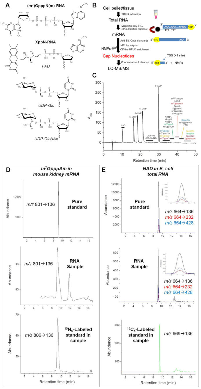
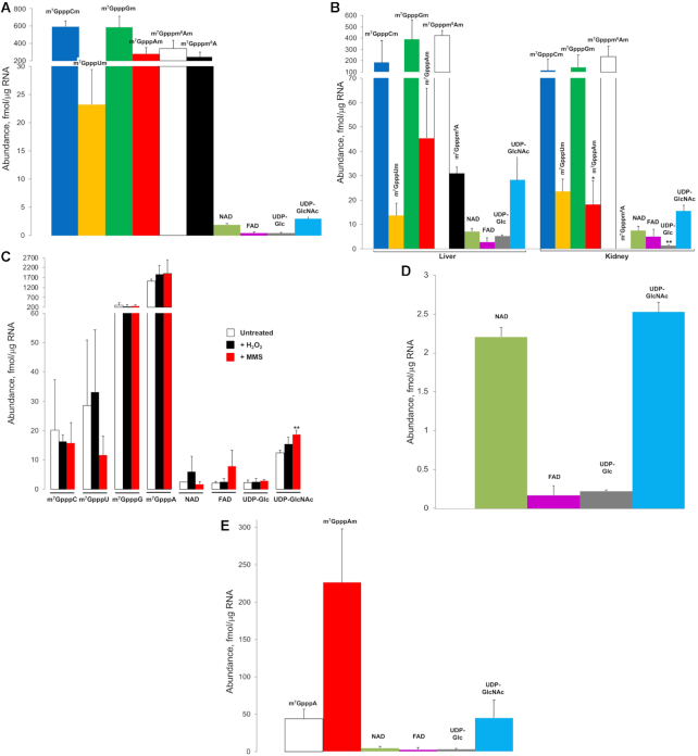
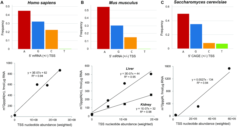
Chemical modification of transcripts with 5' caps occurs in all organisms. Here, we report a systems-level mass spectrometry-based technique, CapQuant, for quantitative analysis of an organism's cap epitranscriptome. The method was piloted with 21 canonical caps-m7GpppN, m7GpppNm, GpppN, GpppNm, and m2,2,7GpppG-and 5 'metabolite' caps-NAD, FAD, UDP-Glc, UDP-GlcNAc, and dpCoA. Applying CapQuant to RNA from purified dengue virus, Escherichia coli, yeast, mouse tissues, and human cells, we discovered new cap structures in humans and mice (FAD, UDP-Glc, UDP-GlcNAc, and m7Gpppm6A), cell- and tissue-specific variations in cap methylation, and high proportions of caps lacking 2'-O-methylation (m7Gpppm6A in mammals, m7GpppA in dengue virus). While substantial Dimroth-induced loss of m1A and m1Am arose with specific RNA processing conditions, human lymphoblast cells showed no detectable m1A or m1Am in caps. CapQuant accurately captured the preference for purine nucleotides at eukaryotic transcription start sites and the correlation between metabolite levels and metabolite caps.
© The Author(s) 2019. Published by Oxford University Press on behalf of Nucleic Acids Research.
Figures
Similar articles
-
A systems-level mass spectrometry-based technique for accurate and sensitive quantification of the RNA cap epitranscriptome.Nat Protoc. 2023 Sep;18(9):2671-2698. doi: 10.1038/s41596-023-00857-0. Epub 2023 Aug 11. Nat Protoc. 2023. PMID: 37567932 Review.
-
The Mysterious World of Non-Canonical Caps - What We Know and Why We Need New Sequencing Techniques.Chembiochem. 2025 Feb 1;26(3):e202400604. doi: 10.1002/cbic.202400604. Epub 2024 Oct 27. Chembiochem. 2025. PMID: 39248054 Free PMC article. Review.
-
Molecular basis for specific viral RNA recognition and 2'-O-ribose methylation by the dengue virus nonstructural protein 5 (NS5).Proc Natl Acad Sci U S A. 2015 Dec 1;112(48):14834-9. doi: 10.1073/pnas.1514978112. Epub 2015 Nov 17. Proc Natl Acad Sci U S A. 2015. PMID: 26578813 Free PMC article.
-
If the 5' cap fits (wear it) - Non-canonical RNA capping.RNA Biol. 2024 Jan;21(1):1-13. doi: 10.1080/15476286.2024.2372138. Epub 2024 Jul 15. RNA Biol. 2024. PMID: 39007883 Free PMC article. Review.
-
Structural insights into dpCoA-RNA decapping by NudC.RNA Biol. 2021 Oct 15;18(sup1):244-253. doi: 10.1080/15476286.2021.1936837. Epub 2021 Jun 18. RNA Biol. 2021. PMID: 34074215 Free PMC article.
Cited by
-
Current landscape of mRNA technologies and delivery systems for new modality therapeutics.J Biomed Sci. 2024 Sep 10;31(1):89. doi: 10.1186/s12929-024-01080-z. J Biomed Sci. 2024. PMID: 39256822 Free PMC article. Review.
-
Nanopore-Based Detection of Viral RNA Modifications.mBio. 2022 Jun 28;13(3):e0370221. doi: 10.1128/mbio.03702-21. Epub 2022 May 17. mBio. 2022. PMID: 35579392 Free PMC article. Review.
-
RIG-I recognizes metabolite-capped RNAs as signaling ligands.Nucleic Acids Res. 2023 Aug 25;51(15):8102-8114. doi: 10.1093/nar/gkad518. Nucleic Acids Res. 2023. PMID: 37326006 Free PMC article.
-
Regulation and functions of non-m6A mRNA modifications.Nat Rev Mol Cell Biol. 2023 Oct;24(10):714-731. doi: 10.1038/s41580-023-00622-x. Epub 2023 Jun 27. Nat Rev Mol Cell Biol. 2023. PMID: 37369853 Review.
-
Toll/interleukin-1 receptor (TIR) domain-containing proteins have NAD-RNA decapping activity.Nat Commun. 2024 Mar 13;15(1):2261. doi: 10.1038/s41467-024-46499-y. Nat Commun. 2024. PMID: 38480720 Free PMC article.
References
-
- Kastern W.H., Berry S.J.. Non-methylated guanosine as 5′ terminus of capped messenger-rna from insect oocytes. Biochem. Biophys. Res. Commun. 1976; 71:37–44. - PubMed
-
- Wei C., Gershowitz A., Moss B.. N6, O2′-dimethyladenosine a novel methylated ribonucleoside next to the 5′ terminal of animal cell and virus mRNAs. Nature. 1975; 257:251–253. - PubMed
Publication types
MeSH terms
Substances
Grants and funding
LinkOut - more resources
Full Text Sources
Other Literature Sources
Molecular Biology Databases
Miscellaneous
