. 2020 Feb 20;22(2):216-228.
doi: 10.1093/neuonc/noz159.

Simultaneous targeting of DNA replication and homologous recombination in glioblastoma with a polyether ionophore

Affiliations

Simultaneous targeting of DNA replication and homologous recombination in glioblastoma with a polyether ionophore

Yi Chieh Lim et al. Neuro Oncol. .

Abstract

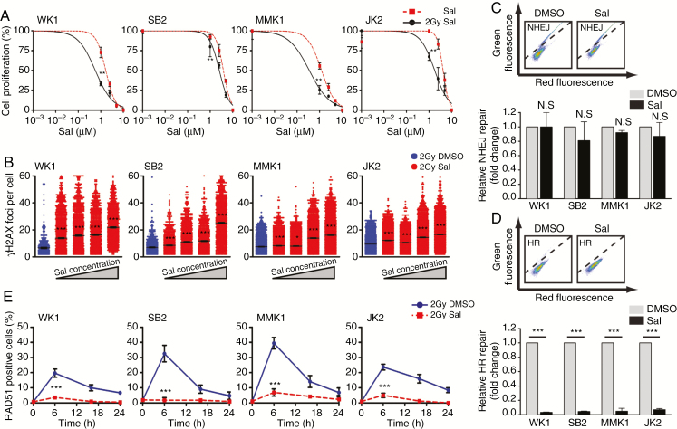
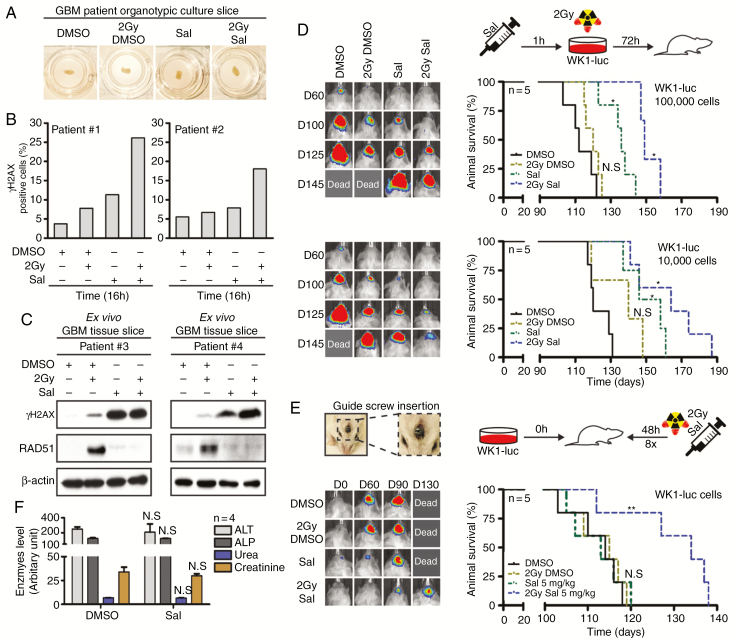
Background: Despite significant endeavor having been applied to identify effective therapies to treat glioblastoma (GBM), survival outcomes remain intractable. The greatest nonsurgical benefit arises from radiotherapy, though tumors typically recur due to robust DNA repair. Patients could therefore benefit from therapies with the potential to prevent DNA repair and synergize with radiotherapy. In this work, we investigated the potential of salinomycin to enhance radiotherapy and further uncover novel dual functions of this ionophore to induce DNA damage and prevent repair.

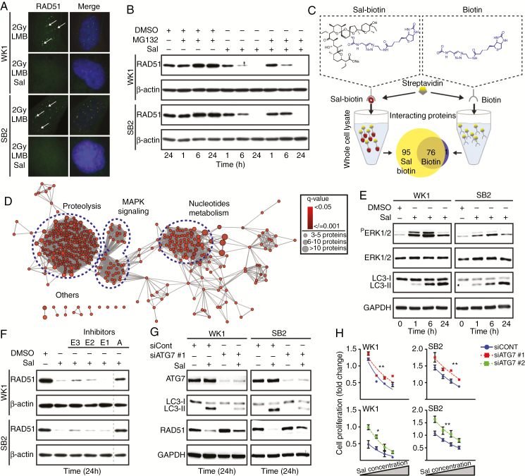
Methods: In vitro primary GBM models and ex vivo GBM patient explants were used to determine the mechanism of action of salinomycin by immunoblot, flow cytometry, immunofluorescence, immunohistochemistry, and mass spectrometry. In vivo efficacy studies were performed using orthotopic GBM animal xenograft models. Salinomycin derivatives were synthesized to increase drug efficacy and explore structure-activity relationships.

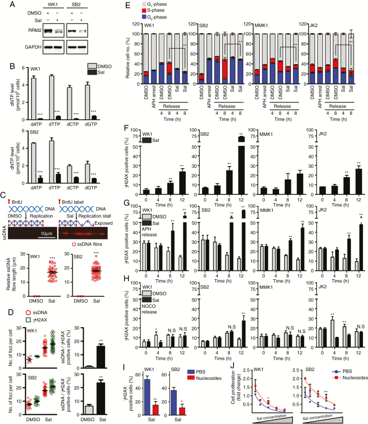
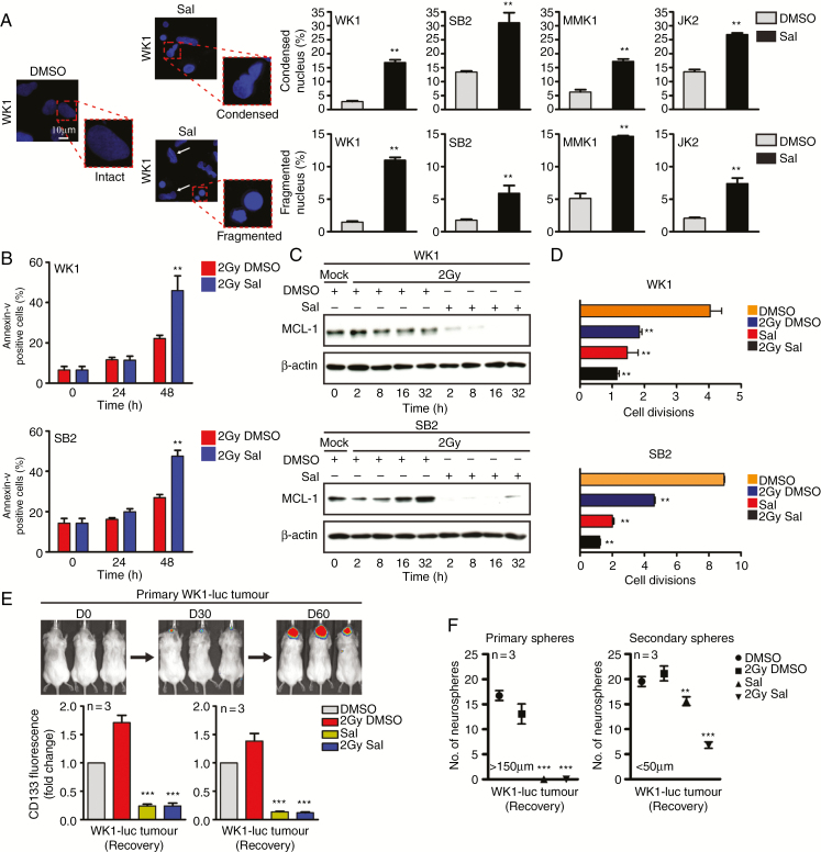
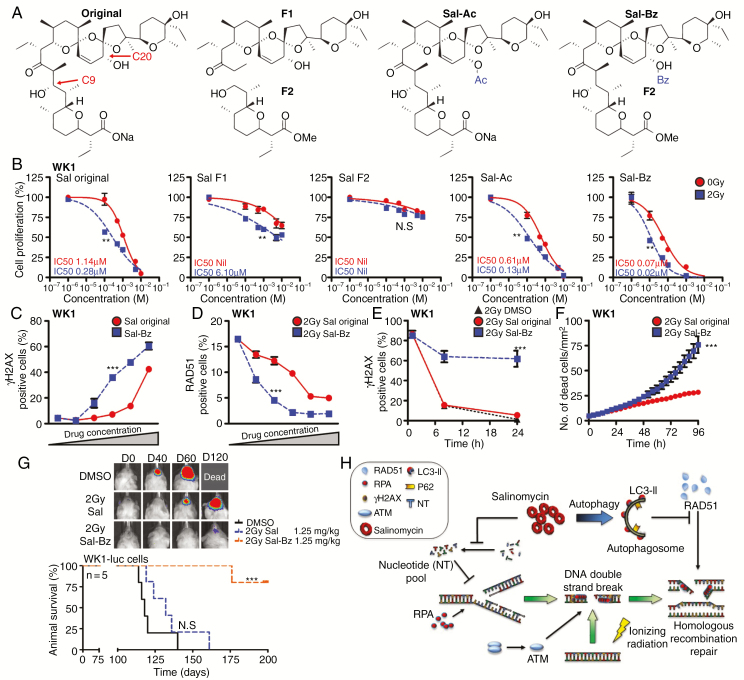
Results: Here we report novel dual functions of salinomycin. Salinomycin induces toxic DNA lesions and prevents subsequent recovery by targeting homologous recombination (HR) repair. Salinomycin appears to target the more radioresistant GBM stem cell-like population and synergizes with radiotherapy to significantly delay tumor formation in vivo. We further developed salinomycin derivatives which display greater efficacy in vivo while retaining the same beneficial mechanisms of action.

Conclusion: Our findings highlight the potential of salinomycin to induce DNA lesions and inhibit HR to greatly enhance the effect of radiotherapy. Importantly, first-generation salinomycin derivatives display greater efficacy and may pave the way for clinical testing of these agents.

Keywords: DNA damage; drug discovery; glioblastoma; homologous recombination.


Figures

Fig. 1
Fig. 1
Salinomycin prevents the resolution of DNA DSBs. (A) Salinomycin (Sal) dose response 72 hours post-treatment ± IR (2 Gy). (B) 24 hours post-Sal (0.5 µM–10 µM) treatment. DNA DSBs induction was assessed by γH2AX foci (green) and nuclear staining (blue). Each dot represents the foci number of individual cells. (C, D) DNA DSB (NHEJ and HR) repair was determined by quantifying GFP positive cells (above diagonal line) at 48 hours post-DMSO or post-Sal treatment (middle). NHEJ and HR was depicted as fold change (bottom). (E) RAD51 positive cells (>5 foci per nucleus) were assessed following DMSO (blue) or Sal (red) treatment (10 µM) followed by IR (2 Gy). Statistical significance: **P < 0.01, ***P < 0.001.
Fig. 2
Fig. 2
Salinomycin induces autophagy to target HR. (A) GNS cells were given LMB (20 nM) to inhibit nuclear protein export followed by DMSO or Sal (10 µM) with IR (2 Gy). Six hours posttreatment RAD51 foci were assessed (white arrows). (B) One hour prior to treatment, GNS cells were given the proteasome inhibitor (MG132, 20 µM) following DMSO or Sal treatment (10 µM). RAD51 protein was determined by immunoblot. (C) Schematic of the label-free quantification approach used for drug–protein interaction study. A Venn diagram depicted the number of interacting proteins with Sal-biotin or biotin (false discovery rate <0.05). (D) The functionally grouped network of proteins that interacted with Sal-biotin. Three proteins per term as nodes linked with ≥0.4 kappa score level. Overrepresented term per group was shown. (E) Immunoblot to validate MAPK and autophagy in Sal-treated cells (10 µM). (F) GNS cells received ubiquitin E3 (PYR-41, 50 µM), E2 (CDC34, 50 µM), E1 (MLN4924, 1 µM) ligases and autophagosome (A) (3-Methyladenine, 5 mM) inhibitors as indicated (+) for one hour prior to Sal treatment (10 µM). The vertical line represents a splice mark between samples from the same gel. (G) RAD51 and LC3 levels were assessed by immunoblot in siControl versus siATG7 knockdown cells. Following siRNA knockdown, cell proliferation was assessed 48 hours post-Sal treatment (2.5 µM–10 µM). Statistical significance: **P < 0.01, ***P < 0.001.
Fig. 3
Fig. 3
Salinomycin causes replication-associated DNA DSBs. (A) RRM2 protein expression was assessed by immunoblot following Sal treatment (10 µM). (B) dNTP level was measured 24 hours post-Sal treatment (10 µM). (C) ssDNA combing strategy used (top) and representative ssDNA fiber (middle). GNS cells were pre-labeled with BrdU for 24 hours prior to DMSO or Sal treatment (10 µM) for 8 hours to determine native ssDNA fibers (bottom). (D) GNS cells were pre-labeled with BrdU prior to DMSO or Sal treatment (10 µM) for 8 hours. Positive cells (%) showing dual ssDNA and γH2AX foci (>5 foci per nucleus, bottom). (E) GNS cells were synchronized in G1/S phase using APH. Cell-cycle analysis was conducted following the release and subsequent DMSO or Sal (10 µM) treatment. (F) Analysis of γH2AX positive cells (>5 foci per nucleus) following Sal treatment (10 µM) in asynchronous cells. (G, H) GNS cells were synchronized using APH (G1/S phase) or NOCO (G2 phase). DNA DSBs were determined by quantifying γH2AX positive cells (>5 foci per nucleus) following Sal treatment (10 µM). (I) GNS cells received PBS or nucleoside supplement (40 µM) prior to Sal treatment (10 µM). γH2AX positive cells (>5 foci per nucleus) were assessed 12 hours post-treatment. (J) Cell proliferation was assessed 48 hours post-Sal treatment (1 µM–10 µM) in PBS versus nucleoside supplemented cells (40 µM). Statistical significance: *P < 0.05, **P < 0.01.
Fig. 4
Fig. 4
Salinomycin targets the radioresistant stem cell–like population. (A) Intact, condensed, and fragmented nuclei (4′,6′-diamidino-2-phenylindole blue) were assessed 24 hours post-DMSO versus post-Sal (10 µM) treatment, quantitation (right). (B) Flow cytometry was performed to determine cell death following Sal treatment (10 µM) with ± IR (2 Gy). (C) Immunoblot was performed to determine MCL-1 protein expression following Sal (10 µM) with ± IR (2 Gy) treatment. (D) CFSE was used to track cell division, GNS cells were treated with Sal (10 µM) ± IR (2 Gy) and allowed to recover for 72 hours prior to labeling. Cell division was assessed 96 hours later. (E, F) WK1-luc tumor cells were isolated from orthotopic xenografts at day 60 and received Sal (10 µM) with ± IR (2 Gy) treatment for 72 hours prior to CD133 flow cytometric expression analysis as indicated; CD133 expression was assessed during treatment (treated) and after Sal had been removed for 96 hours (recovery). Neurosphere formation was assessed 7 days post-treatment withdrawal as indicated. Statistical significance: **P < 0.01, ***P < 0.001.
Fig. 5
Fig. 5
Salinomycin synergizes with IR to prolong survival in vivo. (A) GBM patient organotypic slice cultures were harvested 16 hours post-Sal (10 µM) with ± IR (2 Gy) treatment. (B) IHC was conducted to assess γH2AX positive cells post-treatment (patient specimens #1 and #2) and (C) γH2AX and RAD51 were determined by immunoblot (patient specimens #3 and #4). (D) WK1-luc cells received 72 hours of Sal treatment (10 µM) with ± IR (2 Gy). Dead cells were excluded by trypan blue, 1 × 10⁴ and 1 × 10⁵ WK1-luc cells were engrafted orthotopically into NOD/SCID mice and survival determined by Kaplan–Meier plot. (E) Intracranial studies were conducted using a guide screw approach, a total dose of 40 mg/kg (brain weight) of DMSO versus Sal in 8 fractions of 5 mg/kg was given. (F) Control cull (n = 4) was performed 5 days post-treatment. Serum was collected for the analysis of liver (alanine aminotransferase, alkaline phosphatase, and creatinine) and kidney (urea) injuries. Statistical significance: *P < 0.05, **P < 0.01.
Fig. 6
Fig. 6
Benzene derivative analysis in vitro and in vivo. (A) Diagram showing the generated structural analogues of salinomycin. (B) A dose response curve of cell proliferation was used to compare derivatives versus Sal 96 hours posttreatment. (C, D) γH2AX or RAD51 positive cells (>5 foci per cell) were assessed by immunofluorescence to compare the response between Sal and Sal-Bz (1 µM–10 µM). (E, F) GNS cells were pretreated for one hour with 1 µM of DMSO, Sal, or Sal-Bz in combination with IR (2 Gy). γH2AX positive cells and cell death were quantified using IncuCyte analysis. (G) WK1-luc cells were intracranially engrafted in NOD/RAG mice; animals received a total dose of 10 mg/kg (brain weight) of DMSO, Sal, or Sal-BZ in 8 fractions of 1.25 mg/kg and survival determined by Kaplan–Meier plot. Statistical significance: **P < 0.01, ***P < 0.001. (H) Simplified model of the mechanism of action of salinomycin.

Comment in

References

    1. Hegi ME, Diserens AC, Gorlia T, et al. MGMT gene silencing and benefit from temozolomide in glioblastoma. N Engl J Med. 2005;352(10):997–1003. - PubMed
    1. Bao S, Wu Q, McLendon RE, et al. Glioma stem cells promote radioresistance by preferential activation of the DNA damage response. Nature. 2006;444(7120):756–760. - PubMed
    1. Lim YC, Roberts TL, Day BW, et al. Increased sensitivity to ionizing radiation by targeting the homologous recombination pathway in glioma initiating cells. Mol Oncol. 2014;8(8):1603–1615. - PMC - PubMed
    1. Short SC, Martindale C, Bourne S, Brand G, Woodcock M, Johnston P. DNA repair after irradiation in glioma cells and normal human astrocytes. Neuro Oncol. 2007;9(4):404–411. - PMC - PubMed
    1. Mao Z, Bozzella M, Seluanov A, Gorbunova V. DNA repair by nonhomologous end joining and homologous recombination during cell cycle in human cells. Cell Cycle. 2008;7(18):2902–2906. - PMC - PubMed

Publication types